. 2011 Jan;155(1):112-22.
doi: 10.1016/j.virusres.2010.09.007. Epub 2010 Sep 21.

Characterization of Durham virus, a novel rhabdovirus that encodes both a C and SH protein

Affiliations

Characterization of Durham virus, a novel rhabdovirus that encodes both a C and SH protein

A B Allison et al. Virus Res. 2011 Jan.

Abstract

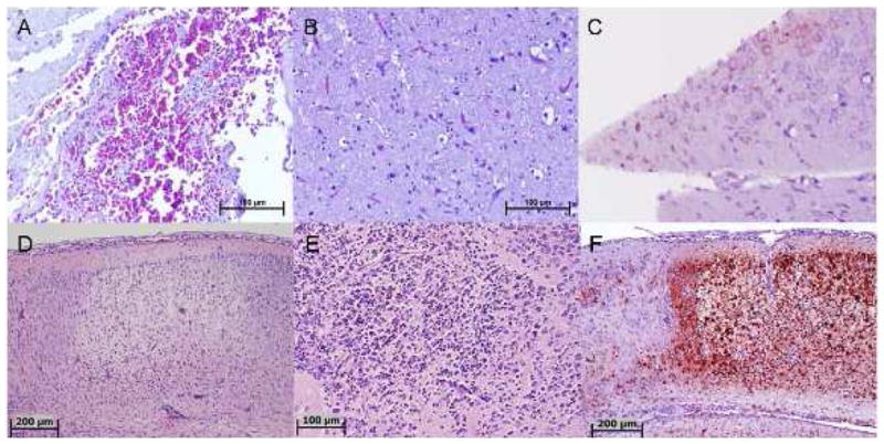
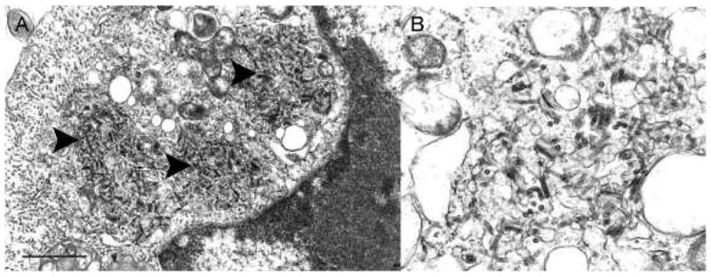
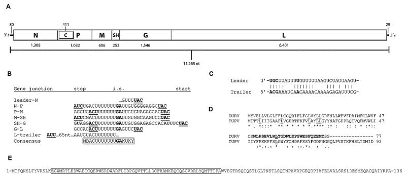
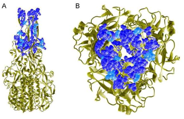
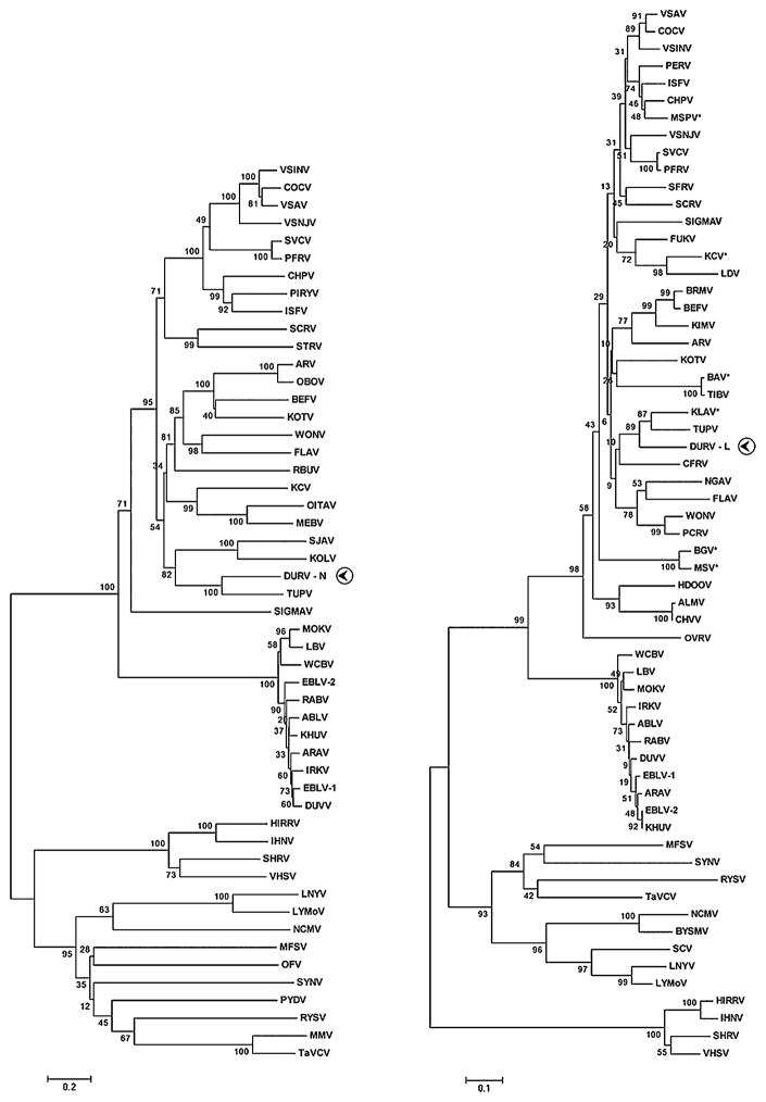
The family Rhabdoviridae is a diverse group of non-segmented, negative-sense RNA viruses that are distributed worldwide and infect a wide range of hosts including vertebrates, invertebrates, and plants. Of the 114 currently recognized vertebrate rhabdoviruses, relatively few have been well characterized at both the antigenic and genetic level; hence, the phylogenetic relationships between many of the vertebrate rhabdoviruses remain unknown. The present report describes a novel rhabdovirus isolated from the brain of a moribund American coot (Fulica americana) that exhibited neurological signs when found in Durham County, North Carolina, in 2005. Antigenic characterization of the virus revealed that it was serologically unrelated to 68 other known vertebrate rhabdoviruses. Genomic sequencing of the virus indicated that it shared the highest identity to Tupaia rhabdovirus (TUPV), and as only previously observed in TUPV, the genome encoded a putative C protein in an overlapping open reading frame (ORF) of the phosphoprotein gene and a small hydrophobic (SH) protein located in a novel ORF between the matrix and glycoprotein genes. Phylogenetic analysis of partial amino acid sequences of the nucleoprotein and polymerase protein indicated that, in addition to TUPV, the virus was most closely related to avian and small mammal rhabdoviruses from Africa and North America. In this report, we present the morphological, pathological, antigenic, and genetic characterization of the new virus, tentatively named Durham virus (DURV), and discuss its potential evolutionary relationship to other vertebrate rhabdoviruses.


Figures

Fig. 1
Fig. 1
Pathology of DURV in avian and mammalian brain sections. (A-C): Sections of coot brain naturally-infected with DURV. (A) Severe congestion in the meninges (H&E); (B) Diffuse neuronal degeneration (accompanied by prominent vacuolation), mononuclear inflammatory cellular infiltration, and prominent capillary vessels (H&E); (C) Immunohistochemical staining demonstrating neurons with perinuclear DURV antigen. (D-F): Sections of mouse brain experimentally-infected with DURV. (D) Neurophil and neurons of the cerebral cortex; inflammatory cellular infiltrate has started from the bottom edge of the lesions. A prominent reactive blood vessel is visible (bottom) and marked meningitis is also present (top) (H&E); (E) A necrotic focus in the deeper subcortical nuclei showing loss of neurons and infiltration with many neutrophils and macrophages (H&E); (F) Immunohistochemical staining of same area of brain as shown in (D), demonstrating necrosis of the central cortex, with strong staining for DURV antigen. Scattered neurons in the adjacent area (far left) are also positive for viral antigen.
Fig. 2
Fig. 2
Ultrastructure of DURV in ultrathin sections of Vero cells. (A) Portion of a cell cytoplasm with three areas of virus formation (arrowheads); Bar = 1 μm. (B) Detail of a virus formation area, demonstrating virions budding into enlarged endoplasmic reticulum cisterns; Bar = 100 nm.
Fig. 3
Fig. 3
DURV genome and regions of interest. (A) Schematic organization of the DURV genome. Including the 3′ leader and 5′ trailer sequences, the DURV genome was 11,265 nucleotides (nt) in length. The length of each gene, in nt, encoding the N, P, M, SH, G, and L proteins, is listed below the schematic. The length of the C gene (encoded in an overlapping reading frame of the P gene), along with the leader and trailer sequences, are noted above the genome schematic. (B) Transcription initiation, intergenic, and transcription termination/polyadenylation sequences. The start and stop codon for each gene is underlined and in bold. The intergenic sequence (i.s.) between each transcription unit, where applicable, is in bold. The consensus sequence for the transcription termination, intergenic (in bold), and transcription initiation regions is boxed. (C) Complementarity of the 3′ leader and 5′ trailer sequences, showing a 65% (19/29nt) inverse identity. Nucleotides that are conserved among the vertebrate rhabdoviruses (positions 1–3; 10) are in bold. (D) Amino acid alignment of the SH proteins of DURV and TUPV. Asterisks denote identity; colons and periods represent conserved and semi-conserved amino acid substitutions, respectively. Conserved leucine residues are underlined. The C-terminal region of the SH protein of DURV that shares homology to the α1 and α2 regions of the SAM domain of the EphA3 protein tyrosine kinase receptor is shown in bold. (E) Amino acid sequence of the DURV C protein. The region of the C protein that shows homology to the fusion domain of the VSINV G protein, as well as to the dynein heavy chains of Culex quinquefasciatus and Aedes aegypti, is boxed.
Fig. 4
Fig. 4
DURV C protein identity and homology to the VSINV G protein. The ribbon schematic of the post-fusion homotrimeric form of the G protein of VSINV (PDB 2CMZ) is shown in tan. The region of the VSINV G protein (amino acids 55–103) which aligns with the DURV C protein (amino acids 15–71) is rendered as van der Waals space-filling and corresponds to domain IV, the fusion domain of the VSINV G protein. The 15 amino acid residues of the DURV C protein which are identical to the VSINV G protein are shown in dark blue, while the five amino acid substitutions that are conserved are in light blue. Panel A shows a side view of the VSINV G protein with the distal ends of the fusion loops pointing upwards, while panel B is an enlarged 90° downward rotation of the fusion loops.
Figure 5
Figure 5
Evolutionary relationships of DURV with representative rhabdoviruses generated by neighbor-joining phylogenies of the N and L proteins. DURV sequences and the corresponding protein used in the phylogeny are indicated with a circled arrowhead. Asterisks beside ICTV abbreviations in the L tree denote new sequences. Bootstrap values were determined using 2000 replicates and are listed at each node. Branch lengths are drawn to scale. The trees were calculated using Poisson correction and evolutionary distances are represented as the number of amino acid substitutions per site. Gaps in the alignments were analyzed by complete deletion. Amino acid sequences used to construct the N and L trees were: ABLV, Australian bat lyssavirus (N: AAD01267; L: NP_478343); ALMV, Almpiwar virus (L: AAZ43273); ARAV, Aravan virus (N: Q6X1D8; L: ABV03822); ARV, Adelaide River virus (N: Q65111; L: AAG10421); BAV, Bivens Arm virus (L: GU085726); BEFV, Bovine ephemeral fever virus (N: NP_065398; L: NP_065409); BGV, Bahia Grande virus (L: HQ207195); BRMV, Berrimah virus (L: AAZ43265); BYSMV, Barley yellow striate mosaic virus (L: ACT21686); CFRV, China fish rhabdovirus (L: AAX86686); COCV, Cocal virus (N: ACB47434; L: ACB47438); CHPV, Chandipura virus (N: P11211; L: P13179); CHVV, Charleville virus (L: AAZ43300); DURV, Durham virus (FJ952155); DUVV, Duvenhage virus (N: Q66453; L: ABZ81216); EBLV-1, European bat lyssavirus (N: AAX62875; L: ABZ81181); EBLV-2, European bat lyssavirus 2 (N: YP_001285393; L: ABZ81191); FLAV, Flanders virus (N: AAN73283; L: AAN73288); FUKV, Fukuoka virus (L: AAZ43279); HDOOV, Humpty Doo virus (L: AAZ43271); HIRRV, Hirame rhabdovirus (N: ACO87995; L: NP_919035); IHNV, Infectious hematopoietic necrosis virus (N: Q08449; L: CAA52076); IRKV, Irkut virus (N: Q5VKP6; L: ABV03823); ISFV, Isfahan virus (N: Q5K2K7; L: Q5K2K3); KCV, Kern Canyon virus (N: ABE69215; L: HQ207197); KHUV, Khujand virus (N: Q6X1D4; L: ABV03824); KIMV, Kimberley virus (L: AAZ43266); KLAV, Klamath virus (L: GU085725); KOLV, Kolongo virus (N: ABE69214); KOTV, Kotonkon virus (N: ABE69213; L: AAZ43267); LBV, Lagos bat virus (N: ABF56214; L: ABZ81171); LDV, Le Dantec virus (L: AAZ43278); LNYV, Lettuce necrotic yellows virus (N: YP_425087; L: YP_425092); LYMoV, Lettuce yellow mottle virus (N: YP_002308371; L: YP_002308376); MEBV, Mount Elgon bat virus (N: ABE69217); MFSV, Maize fine streak virus (N: YP_052843; L: YP_052849); MMV, Maize mosaic virus (N: YP_052850; L: YP_052855); MOKV, Mokola virus (N: YP_142350; L: ABZ81211); MSPV, Malpais Springs virus (L: GU085727); MSV, Muir Springs virus (L: HQ207196); NCMV, Northern cereal mosaic virus (N: NP_057954; L: NP_597914); NGAV, Ngaingan virus (L: AAZ43277); OBOV, Obodhiang virus (N: ABE69212); OFV, Orchid fleck virus (N: BAH97109; L: YP_001294929); OITAV, Oita virus (N: BAD13431); OVRV: Oak Vale virus (L: AAZ43298); PCRV, Parry Creek virus (L: AAZ43275); PERV, Perinet virus (L: AAZ43280); PFRV, Pike fry rhabdovirus (N: ACP27998; L: ACP28002); PIRYV, Piry virus (N: P26037); PYDV, Potato yellow dwarf virus (N: ABW35154); RABV, Rabies virus (N: ACN51666; L: Q66T60); RBUV, Rochambeau virus (N: ABE69218); RYSV, Rice yellow stunt virus (N: NP_620496; L: NP_620502); SCRV, Siniperca chuatsi rhabdovirus (N: YP_802937; L: YP_802942); SCV, Strawberry crinkle virus (L: AAP03645); SFRV, Starry flounder rhabdovirus (L: AAS02285); SHRV, Snakehead rhabdovirus (N: NP_050580; L: NP_050585); SIGMAV, Sigma virus (N: ACV67011; L: ACU65438); SJAV, Sandjimba virus (N: ABE69216); STRV, Sea trout rhabdovirus (N: AAL35756); SVCV, Spring viremia of carp virus (N: ABW24033; L: Q91DR9); SYNV, Sonchus yellow net virus (N: P10550; L: NP_042286); TaVCV, Taro vein chlorosis virus (N: YP_224078; L: YP_224083); TIBV, Tibrogargan virus (L: AAZ43274); TUPV, Tupaia rhabdovirus (N: YP_238528; L: YP_238534); VHSV, Viral hemorrhagic septicemia virus (N: P24378; L: CAB40833); VSINV, Vesicular stomatitis Indiana virus (N: P11212; L: NP_041716); VSAV, Vesicular stomatitis Alagoas virus (N: ACB47439; L: ACB47443); VSNJV, Vesicular stomatitis New Jersey virus (N: P04881; L: P16379); WCBV, West Caucasian bat virus (N: Q5VKP2; L: ABV03821); WONV, Wongabel virus (N: YP_002333271; L: AAZ43276).

References

    1. Abraham G, Banerjee AK. Sequential transcription of the genes of vesicular stomatitis virus. Proc Natl Acad Sci U S A. 1976;73:1504–1508. - PMC - PubMed
    1. Alonso M, Kim CH, Johnson MC, Pressley M, Leong J. The NV gene of snakehead rhabdovirus (SHRV) is not required for pathogenesis, and a heterologous glycoprotein can be incorporated into the SHRV envelope. J Virol. 2004;78:5875–5882. - PMC - PubMed
    1. Blom N, Gammeltoft S, Brunak S. Sequence- and structure-based prediction of eukaryotic protein phosphorylation sites. J Mol Biol. 1999;294:1351–1362. - PubMed
    1. Bourhy H, Cowley JA, Larrous F, Holmes EC, Walker PG. Phylogenetic relationships among rhabdoviruses inferred using the L polymerase gene. J Gen Virol. 2005;86:2849–2858. - PubMed
    1. Calisher CH, Karabatsos N, Zeller H, Digoutte JP, Tesh RB, Shope RE, Travassos da Rosa AP, St George TD. Antigenic relationships among rhabdoviruses from vertebrates and hematophagous arthropods. Intervirology. 1989;30:241–257. - PubMed

Publication types

MeSH terms

Associated data

LinkOut - more resources