. 2010 Oct;18 Suppl 3(Suppl 3):S35-52.
doi: 10.1016/j.joca.2010.04.015.

The OARSI histopathology initiative - recommendations for histological assessments of osteoarthritis in the guinea pig

Affiliations

The OARSI histopathology initiative - recommendations for histological assessments of osteoarthritis in the guinea pig

V B Kraus et al. Osteoarthritis Cartilage. 2010 Oct.

Abstract

Objective: This review focuses on the criteria for assessing osteoarthritis (OA) in the guinea pig at the macroscopic and microscopic levels, and recommends particular assessment criteria to assist standardization in the conduct and reporting of preclinical trails in guinea pig models of OA.

Methods: A review was conducted of all OA studies from 1958 until the present that utilized the guinea pig. The PubMed database was originally searched August 1, 2006 using the following search terms: guinea pig and OA. We continued to check the database periodically throughout the process of preparing this chapter and the final search was conducted January 7, 2009. Additional studies were found in a review of abstracts from the OsteoArthritis Research Society International (OARSI) conferences, Orthopaedic Research Society (ORS) conferences, and literature related to histology in other preclinical models of OA reviewed for relevant references. Studies that described or used systems for guinea pig joint scoring on a macroscopic, microscopic, or ultrastructural basis were included in the final comprehensive summary and review. General recommendations regarding methods of OA assessment in the guinea pig were derived on the basis of a comparison across studies and an inter-rater reliability assessment of the recommended scoring system.

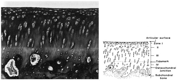
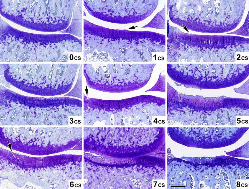
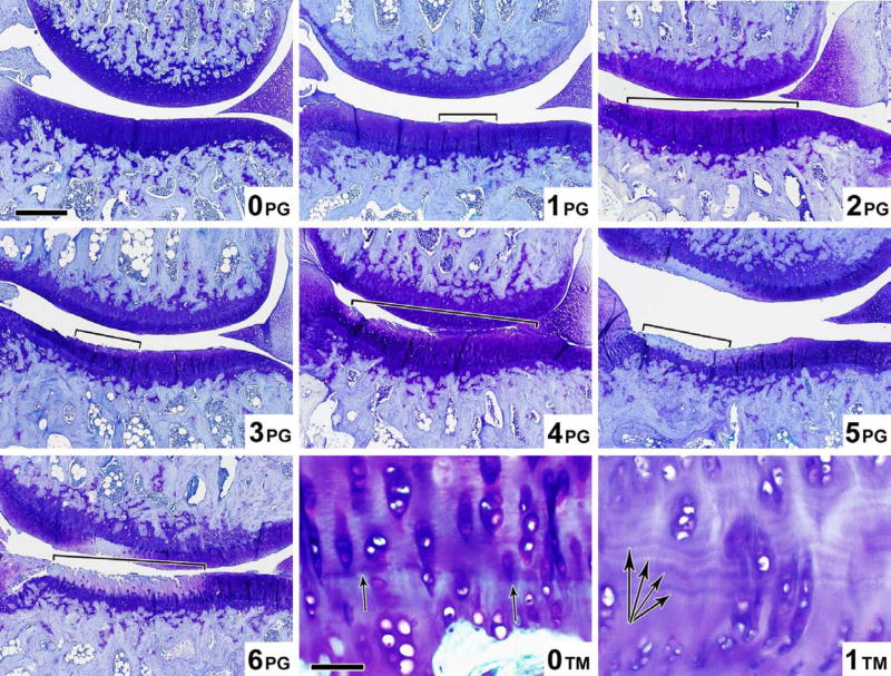
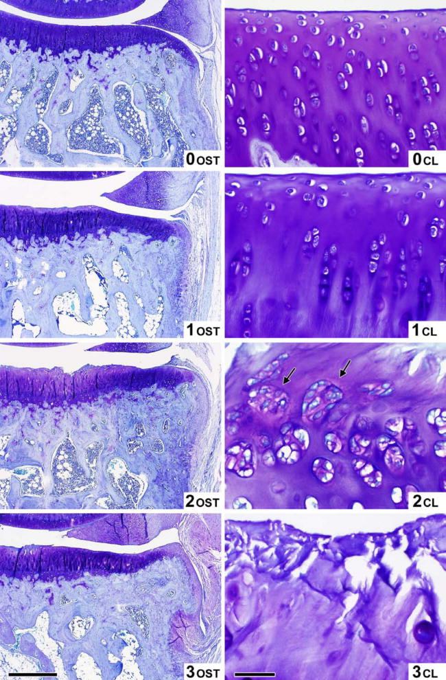
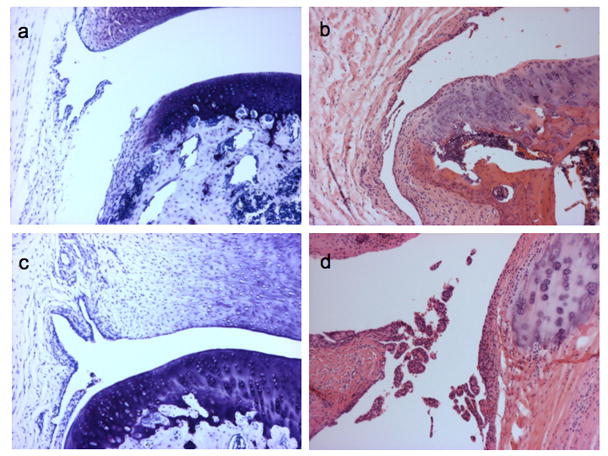
Results: A histochemical-histological scoring system (based on one first introduced by H. Mankin) is recommended for semi-quantitative histological assessment of OA in the guinea pig, due to its already widespread adoption, ease of use, similarity to scoring systems used for OA in humans, its achievable high inter-rater reliability, and its demonstrated correlation with synovial fluid biomarker concentrations. Specific recommendations are also provided for histological scoring of synovitis and scoring of macroscopic lesions of OA.

Conclusions: As summarized herein, a wealth of tools exist to aid both in the semi-quantitative and quantitative assessment of OA in the guinea pig and provide a means of comprehensively characterizing the whole joint organ. In an ongoing effort at standardization, we recommend specific criteria for assessing the guinea pig model of OA as part of an OARSI initiative, termed herein the OARSI-HISTOgp recommendations.


Conflict of interest statement

Conflict of interest statement: No author has any conflict of interest related to this work. The work performed was not influenced at any stage by the support provided.

Figures

Figure 1
Figure 1. Guinea pig knee joint structures
A frontal section through a 12-month-old guinea pig knee joint to demonstrate its structures and the landmarks and features typical of advanced OA at the center of the tibiofemoral joint (M=medial; L=lateral).
Figure 2
Figure 2. The timecourse of spontaneous histological OA in the Hartley guinea pig knee
a) Representative sections of the central medial tibial plateau stained with toluidine blue for each age group (ages 2 to 18 months). Scores of cartilage structure (CS) and proteoglycan content (PG) using the recommended scoring system for the regions shown is as follows: 2 months CS=0/PG=0; 4 months CS=2/PG=1; 7 months CS=5/PG=4; 10 months CS=5/PG=5; 12 months CS=8/PG=5; and 18 months CS=8/PG=6. b) Mean histological scores of the knee demonstrating increasing OA severity with age in this model system (N=6 for each group ages 2–12 months and N=10 for 18 month age group). Error bars = SD.
Figure 3
Figure 3. Macroscopic images of the guinea pig knee joint
a) Guinea pig knee joint demonstrating normal dimensions of approximately 1 cm x 1 cm (width x depth); b) Arial views of the tibial plateau oriented (medial on left, lateral on right) with macroscopic lesions of increasing severity from no lesions (left panel), to moderate surface fissuring (middle panel), and severe full depth erosion to bone (right panel); the diseased areas in the middle and right panels are indicated by the dotted demarcated region.
Figure 4
Figure 4. Landmarks of the central region of the guinea pig tibiofemoral joint
a) A schematic representation of a centrally sectioned guinea pig knee joint based upon Cashin [35] and reproduced by permission of Oxford University Press; b) A series of 4 sections taken at 200 μm intervals in the coronal plane, spanning the anterior of the joint through to the center. Note the elongated appearance of the lateral meniscus (on left) and the more rounded appearance of the medial meniscus (on right) when the center of the knee joint is attained.
Figure 5
Figure 5. Plane of sectioning of the guinea pig knee joint
The lateral aspect of the guinea pig knee showing the plane of sectioning (A-A′) based on Cashin [35] and reproduced by permission of Oxford University Press. Note that the knee is in slight flexion.
Figure 6
Figure 6. Zones of articular cartilage
Accurate scoring requires the ability to distinguish the four zones of articular cartilage as depicted here: I-superficial zone, II-middle zone, III-deep zone, and IV-calcified cartilage zone, and the tidemark separating zones III and IV. Reproduced by permission of Dr. James Williams.
Figure 7
Figure 7. Representative images of guinea pig knees (aged 2–18 months) demonstrating varying severities of histological lesions
Examples of lesions scored by the recommended histochemical-histological scoring scheme. The mean score generated by two experts are shown in the bottom right of each panel in Figures 7a-c. Figure 7a. Cartilage structure (CS) scoring atlas. Microscopic toluidine blue stained images of the guinea pig knee joint demonstrating the 9 levels (scores 0–8) of cartilage structure (CS) corresponding to the criteria in Table 4. Magnification bar 500 μm. Figure 7b. Proteoglycan content (PG) and Tidemark™ scoring atlas. Microscopic toluidine blue stained images of the guinea pig knee joint demonstrating the 7 levels (scores 0–6) of proteoglycan content (PG) (Magnification bar = 500 μm), and the 2 levels (scores 0–1) of Tidemark ™ integrity (Magnification bar = 50 μm) corresponding to the criteria in Table 4. Figure 7c. Osteophyte (OST) and Cellularity (CL) scoring atlas. Microscopic toluidine blue stained images of the guinea pig knee joint demonstrating the 4 levels (scores 0–3) of osteophyte (OST) (Magnification bar = 250 μm), and the 4 levels (scores 0–3) of Cellularity (CL) (Magnification bar = 50 μm) corresponding to the criteria in Table 4.
Figure 8
Figure 8. Examples of synovitis associated with spontaneous OA in the Hartley guinea pig knee joint
Representative images (100x) of synovitis scored according to the recommended scoring system in Table 6. Sections stained with toluidine blue (a and c) or Hematoxylin and Eosin (b and d). Normal synovium from a 60 day old animal (a); Synovitis scores of 3 in a 145 day old animal (b) and a 180 day old animal (c) based on scoring synovial hyperplasia=1 (3–5 layers), villous hyperplasia=1, and scattered infiltrate=1. Synovitis score of 7 in a 2 year old animal (d) (100x) based on scoring synovial hyperplasia=2 (≥6 layers), villous hyperplasia=3, and infiltrate=2.
Figure 9
Figure 9
Effect of caging on guinea pig knee joint histological OA severity. Animals were housed either singly (n=26, white bars), or in pairs (n=14, dark bars) from 2 to 12 months of age. Paraffin central sections of these knee joints (aged 12 months) were scored using the recommended scoring scheme to assess cartilage structure (CS) and proteoglycan content (PG) or a combination of both (Total), of the medial tibia (MT) or entire medial compartment (MED). A comparison of paired housing with single housing by the non-parametric Mann Whitney test, was associated with slight but significantly greater histological scores in the paired-housed animals compared with single-housed animals (*p=0.02; § p =0.04, respectively). Error bars = SEM.

Similar articles

Cited by

References

    1. Silverstein E, Sokoloff L. Natural history of degenerative joint disease in small animals. 5. Osteoarthritis in guinea pigs. Arthritis Rheum. 1958;1:82–86. - PubMed
    1. Bendele AM. Progressive chronic osteoarthritis in femorotibial joints of partial medial meniscectomized guinea pigs. Vet Pathol. 1987;24:444–448. - PubMed
    1. Bendele A, Hulman J. Spontaneous cartilage degeneration in guinea pigs. Arthritis Rheum. 1988;31:561–565. - PubMed
    1. Bendele AM, White SL, Hulman JF. Osteoarthritis in guinea pigs: histopathologic and scanning electron microscopic features. Lab Animal Sci. 1989;39:115–121. - PubMed
    1. Meacock SC, Bodmer JL, Billingham ME. Experimental osteoarthritis in guinea pigs. J Exp Pathol (Oxford) 1990;71:279–293. - PMC - PubMed

Publication types