. 2010 Nov 19;285(47):36625-34.
doi: 10.1074/jbc.M110.159954. Epub 2010 Sep 23.

The dopamine D1-D2 receptor heteromer localizes in dynorphin/enkephalin neurons: increased high affinity state following amphetamine and in schizophrenia

Affiliations

The dopamine D1-D2 receptor heteromer localizes in dynorphin/enkephalin neurons: increased high affinity state following amphetamine and in schizophrenia

Melissa L Perreault et al. J Biol Chem. .

Abstract

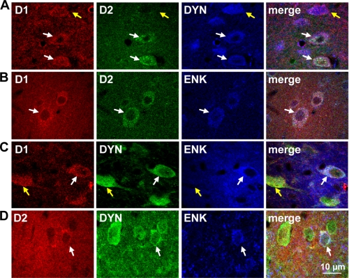
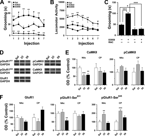
The distribution and function of neurons coexpressing the dopamine D1 and D2 receptors in the basal ganglia and mesolimbic system are unknown. We found a subset of medium spiny neurons coexpressing D1 and D2 receptors in varying densities throughout the basal ganglia, with the highest incidence in nucleus accumbens and globus pallidus and the lowest incidence in caudate putamen. These receptors formed D1-D2 receptor heteromers that were localized to cell bodies and presynaptic terminals. In rats, selective activation of D1-D2 heteromers increased grooming behavior and attenuated AMPA receptor GluR1 phosphorylation by calcium/calmodulin kinase IIα in nucleus accumbens, implying a role in reward pathways. D1-D2 heteromer sensitivity and functional activity was up-regulated in rat striatum by chronic amphetamine treatment and in globus pallidus from schizophrenia patients, indicating that the dopamine D1-D2 heteromer may contribute to psychopathologies of drug abuse, schizophrenia, or other disorders involving elevated dopamine transmission.


Figures

FIGURE 1.
FIGURE 1.
Control data depicting antibody specificity for D1 and D2 receptor coexpression and FRET analysis. A, confocal images revealed no reactivity of the D1-Alexa Fluor 488 antibody in D1R gene-deleted mouse CP. Similarly, the D2-Alexa Fluor 350 antibody did not exhibit reactivity in CP tissue of D2R gene-deleted mice. Note the high level of dendritic staining in this region. B, there was no cross-reactivity of the antibody-linked secondary fluorophores when coincubated with striatal tissue. Emission was detected only when excitation occurred at the appropriate wavelength, with no spectral bleed-through. C, D1R and D2R antibody specificity was further shown by labeling of cells coexpressing both receptors (white arrows) shown together with neurons expressing only the D1R within the same section of NAc shell. D and E, confocal FRET images of a neonatal striatal neuron or in NAc. In both the dendrites (D, inset) and cell bodies, the FRET signal occurred only when the donor (Alexa Fluor 350) and acceptor (Alexa Fluor 488) were present together (white arrows). When either the donor or acceptor was absent, no FRET signal was generated (blue arrows).
FIGURE 2.
FIGURE 2.
Colocalization of the D1 and D2 receptor with dynorphin and enkephalin in rat nucleus accumbens. A and B, confocal images revealed D1R and D2R colocalization with DYN or with ENK (white arrows) in NAc. Neurons that expressed only D1R were for positive for DYN and neurons expressing only D2R were positive for ENK (yellow arrows). C and D, DYN+-ENK+-coexpressing neurons also expressed D1R or D2R (white arrows) in NAc. D1R was not expressed in DYN/ENK+ neurons, nor was D2R expressed in DYN+/ENK cells (yellow arrows).
FIGURE 3.
FIGURE 3.
Colocalization of the D1 and D2 receptor with dynorphin and enkephalin in rat caudate putamen. A and B, confocal images showed that the D1R and D2R were colocalized with DYN or with ENK in CP (white arrows). C and D, neurons coexpressing DYN and ENK also expressed D1R or D2R (white arrows). DYN and ENK also exhibited coexpression in the absence of D1R (yellow arrows). E and F, neurons that expressed only D1R were positive for DYN, and neurons expressing only D2R were positive for ENK (yellow arrows).
FIGURE 4.
FIGURE 4.
The D1 and D2 receptor colocalized in basal ganglia and formed D1-D2 receptor heteromers. A, quantification of the number of D1R-expressing neurons per 300 μm2 area (left panel) and the proportion of D1R-expressing neurons also expressing the D2R (right panel; n = 16–25 sections/region). B, fluorescence from fluorophore-tagged D1R and D2R in neurons coexpressing them showed equal amounts of the receptor in both NAc subregions but differing expression in CP (n = 43–62 neurons/region). AU, arbitrary units. **, p < 0.01, Student's t test. C, example of a representative neuron showing colocalization and interaction (FRET) of D1R and D2R in an NAc core cell body as assessed from individual microdomains (circles). Scale panel denotes FRET efficiency. D, interaction between D1R and D2R as determined by FRET. The FRET signal indicated equivalent FRET efficiencies in NAc core and shell and lower in CP and indicated the presence of D1-D2 heteromers. E, the proportion of D1/D2-coexpressing neurons that exhibited D1-D2 heteromer formation was 91% in NAc and only 24% in CP. Data in bar graphs expressed as mean ± S.E. NAcC, NAc core; NAcS, NAc shell; VP, ventral pallidum; EPN, entopeduncular nucleus.
FIGURE 5.
FIGURE 5.
The D1 and D2 receptor are colocalized on presynaptic terminals in rat nucleus accumbens and caudate putamen but are extrasynaptic in the ventral tegmental area. A–C, the D1R and D2R colocalized with SYN but not PSD95 (white circles) in the NAc core, shell, and CP. D, colocalization of D1R and D2R in the neuropil of the VTA was scarce and was not observed with SYN or PSD95 (white circles). In all regions, individual D1R colocalized with SYN or PSD95 (yellow circles). Individual D2R also showed considerable, but diffuse, overlap with SYN or PSD95 (purple color, merge).
FIGURE 6.
FIGURE 6.
Repeated D1-D2 receptor heteromer activation induced grooming in rats and attenuated AMPA GluR1 receptor phosphorylation in nucleus accumbens. A, SKF 83959 significantly increased the amount of time spent grooming compared with saline (Sal) controls, whereas SKF 83822 attenuated it (both drugs administered at 0.4 mg/kg subcutaneously for 7 days). B, SKF 83959 stimulated locomotion to a lesser degree than SKF 83822 (n = 8 rats/group). **, p < 0.05 compared with every other group; *, p < 0.05 compared with controls; #, p < 0.05 compared with the other drug treatment group; repeated measures analysis of variance followed by Student's t test for post hoc comparisons. C, antagonism of the D2 receptor by acute administration of raclopride (RAC) (0.5 mg/kg intraperitoneal) attenuated grooming following three injections of SKF 83959 or saline (n = 6 rats/group). *, p < 0.05; ***, p < 0.001, as indicated. D, representative blots depicting the effects of seven daily injections of SKF 83959 or SKF 83822 on total and phosphorylated CaMKII and GluR1 levels in NAc (n = 8 rats/group). GAPDH was used as a loading control. E, SKF 83959 or SKF 83822 diminished the total expression and had no effect on phosphorylated levels of CaMKII in NAc with no drug effects in CP. F, in NAc, SKF 83959 significantly reduced CaMKII-mediated phosphorylation of GluR1 at Ser831. SKF 83822 elevated protein kinase A-mediated GluR1 phosphorylation at Ser845 in both NAc and CP. **, p < 0.01; ***, p < 0.001. Data were expressed as mean ± S.E. data in bar graphs analyzed by analysis of variance followed by post-hoc comparisons by Student's t test.
FIGURE 7.
FIGURE 7.
Modulation of D1-D2 receptor heteromer activity in rat striatum by repeated amphetamine treatment and withdrawal. A, representative competition binding curves of [3H]raclopride binding by quinpirole in rat striatum are shown after AMPH (2.5 mg/kg, intraperitoneally) for 5 days or withdrawal for 21 days. The affinity of the D2 receptor for quinpirole was increased by ∼10-fold. The proportion of D2 receptors in the D2high in striatum was increased by AMPH compared with control (VEH), an effect that was maintained following drug withdrawal (WD) (bar graphs, n = 5–7 rats/group). B, representative competition binding curves of [3H]raclopride binding by SKF 83959 in rat striatum are shown after AMPH for 5 days or withdrawal for 21 days. The affinity of the D2 receptor within the D1-D2 heteromer for SKF 83959 was increased by ∼10-fold. The proportion of heteromeric D2high in striatum was elevated by AMPH compared with control (VEH), and this increase in D2high was still present after drug withdrawal (bar graphs) (n = 5–7 rats/group). C, percent stimulation (above baseline) of [35S]GTPγS binding to striatal membranes by dopamine or SKF 83959. AMPH enhanced dopamine-induced [35S]GTPγS incorporation and sensitized [35S]GTPγS incorporation by ∼100-fold in rat striatum after 5 days of treatment. This effect was absent after 21 days of withdrawal. D1-D2 heteromer activity was also markedly enhanced as indicated by rat striatal SKF 83959-induced [35S]GTPγS incorporation with a 100-fold increase in sensitivity. D, increased interaction (FRET) between D1 and D2 receptors in NAc, but not CP, following AMPH treatment (n = 15–22 neurons/region). Data are expressed as percent control. *, p < 0.05; **, p < 0.01; Student's t test.
FIGURE 8.
FIGURE 8.
Increased agonist affinity of D1-D2 receptor heteromer in schizophrenia globus pallidus. A, representative competition binding curves of [3H]raclopride displaced with SKF 83959 in GP of individuals with schizophrenia showing increased affinity of the drug for the D2 receptor within the D1-D2 heteromer. B, the proportion of striatal D2 receptors in the high affinity state (D2high) within the D1-D2 heteromer was significantly higher in schizophrenia brain GP than in control tissue (schizophrenia (SCHIZ), D2high 49.1 ± 4.0%; control (CONT), D2high 28.5 ± 3.5%; n = 4–5/group). **, p < 0.01; Student's t test.

References

    1. Reiner A., Medina L., Haber S. N. (1999) Neuroscience 88, 775–793 - PubMed
    1. Steiner H., Gerfen C. R. (1998) Exp. Brain Res. 123, 60–76 - PubMed
    1. Bertran-Gonzalez J., Bosch C., Maroteaux M., Matamales M., Hervé D., Valjent E., Girault J. A. (2008) J. Neurosci. 28, 5671–5685 - PMC - PubMed
    1. Lee K. W., Kim Y., Kim A. M., Helmin K., Nairn A. C., Greengard P. (2006) Proc. Natl. Acad. Sci. U.S.A. 103, 3399–3404 - PMC - PubMed
    1. Gerfen C. R., Engber T. M., Mahan L. C., Susel Z., Chase T. N., Monsma F. J., Jr., Sibley D. R. (1990) Science 250, 1429–1432 - PubMed

Publication types

MeSH terms