. 2010 Sep 23;467(7314):426-9.
doi: 10.1038/nature09415.

Gut inflammation provides a respiratory electron acceptor for Salmonella

Affiliations

Gut inflammation provides a respiratory electron acceptor for Salmonella

Sebastian E Winter et al. Nature. .

Abstract

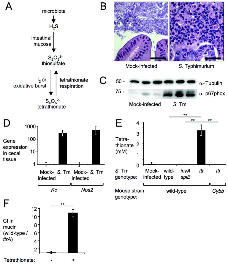
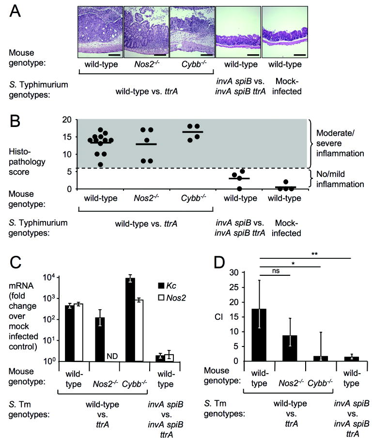
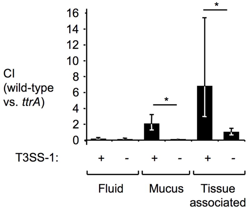
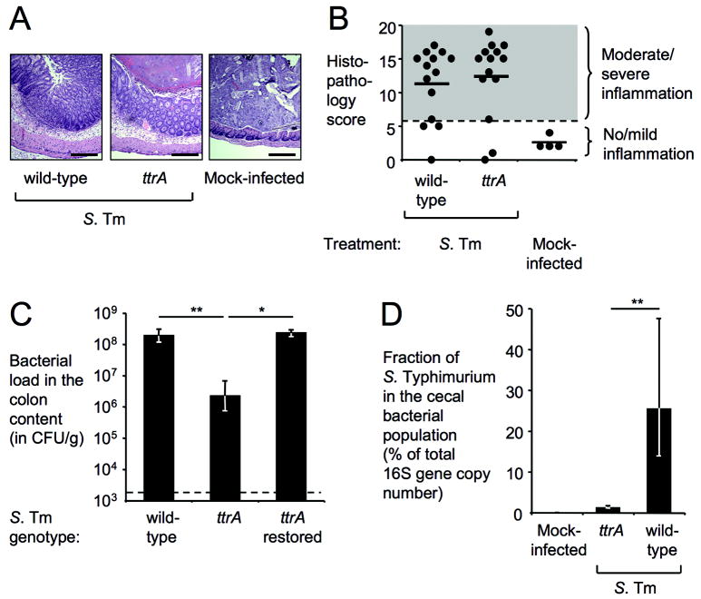
Salmonella enterica serotype Typhimurium (S. Typhimurium) causes acute gut inflammation by using its virulence factors to invade the intestinal epithelium and survive in mucosal macrophages. The inflammatory response enhances the transmission success of S. Typhimurium by promoting its outgrowth in the gut lumen through unknown mechanisms. Here we show that reactive oxygen species generated during inflammation react with endogenous, luminal sulphur compounds (thiosulphate) to form a new respiratory electron acceptor, tetrathionate. The genes conferring the ability to use tetrathionate as an electron acceptor produce a growth advantage for S. Typhimurium over the competing microbiota in the lumen of the inflamed gut. We conclude that S. Typhimurium virulence factors induce host-driven production of a new electron acceptor that allows the pathogen to use respiration to compete with fermenting gut microbes. Thus the ability to trigger intestinal inflammation is crucial for the biology of this diarrhoeal pathogen.


Conflict of interest statement

Competing financial Interests The authors declare no competing financial interests

Figures

Figure 1
Figure 1. Tetrathionate becomes available during inflammation
(A) Schematic of intestinal sulfur metabolism. (B-E) Samples from a mouse colitis model four days after infection with S. Typhimurium (S. Tm) or mock-infection. (B) H&E stained cecal sections. Scale bar, 100 μm. (C) Detection of NADPH oxidase (α–p67phox) or tubulin (α–tubulin) in cecal extracts (n=3). (D) Expression of Kc and Nos2 in cecal RNA samples (n≥3) using qRT-PCR (fold-increases over mock-infection). (E) Tetrathionate detected in cecal contents using LC-MS (n≥3). (F) Competitive indices (CI) for anaerobic growth in mucin broth with (+) or without (-) tetrathionate (n=3). (D-F) Bars represent geometric means ± standard error. **, P < 0.01.
Figure 2
Figure 2. Tetrathionate respiration confers growth advantage
(A-D) Samples from a mouse colitis model (n indicated in B) four days after infection with S. Typhimurium (S. Tm) or mock-infection. (A) H&E stained cecal sections. Scale bar, 400 μm. (B) Blinded histopathology scoring showing averages (bars) and individual scores (circles). (C) Kc (closed bars) and Nos2 (open bars) expression in cecal RNA samples using qRT-PCR (fold-increases over mock-infection). (D) Competitive indices (CI) of indicated S. Typhimurium strains determined by recovering bacteria from colon contents. (C-D) Bars represent geometric means ± standard error. *, P < 0.05; **, P < 0.01; ns, not significant, ND, not determined.
Figure 3
Figure 3. Tetrathionate respiration promotes growth of S. Typhimurium in close proximity to the mucosal surface
Bovine ligated ileal loops (n=3 animals) were infected with a mixture of S. Typhimurium T3SS-1 proficient (+) strains (wild-type [AJB715] versus ttrA mutant[SW661]) or T3SS-1-deficient (-) strains (invA mutant [SW737] versus invA ttrA mutant [SW736]) and samples collected 8 hours after infection from the luminal fluid, mucus scrapings and tissue punches (tissue-associated bacteria). Bars represent geometric means ± standard error. *, P ≤ 0.05.
Figure 4
Figure 4. Tetrathionate respiration increases the abundance of S. Typhimurium in the intestinal lumen
(A-C) Samples from a mouse colitis model (n indicated in B) four days after infection with S. Typhimurium (S. Tm) or mock-infection. (A) H&E stained cecal sections. Scale bar, 400 μm. (B) Blinded histopathology scoring showing averages (bars) and individual scores (circles). (C) Recovery of S. Typhimurium from colon contents. (D) Fraction of S. Typhimurium as percentage of the cecal bacterial population using 16S rRNA gene qRT-PCR (wild-type n=6, ttrA mutant n=6, mock-infected n=4). (C-D) Bars represent geometric means ± standard error. *, P < 0.05; **, P < 0.01.

Comment in

References

    1. Harris JC, Dupont HL, Hornick RB. Fecal leukocytes in diarrheal illness. Ann Intern Med. 1972;76:697–703. - PubMed
    1. Santos RL, et al. Life in the inflamed intestine, Salmonella style. Trends Microbiol. 2009;17:498–506. - PMC - PubMed
    1. Stecher B, et al. Salmonella enterica serovar typhimurium exploits inflammation to compete with the intestinal microbiota. PLoS Biol. 2007;5:2177–2189. - PMC - PubMed
    1. Barman M, et al. Enteric salmonellosis disrupts the microbial ecology of the murine gastrointestinal tract. Infection and immunity. 2008;76:907–915. - PMC - PubMed
    1. Sekirov I, et al. Antibiotic-induced perturbations of the intestinal microbiota alter host susceptibility to enteric infection. Infection and immunity. 2008;76:4726–4736. - PMC - PubMed

Publication types

MeSH terms