. 2010 Sep 23;467(7314):479-83.
doi: 10.1038/nature09377.

Damage-induced phosphorylation of Sld3 is important to block late origin firing

Affiliations

Damage-induced phosphorylation of Sld3 is important to block late origin firing

Jaime Lopez-Mosqueda et al. Nature. .

Abstract

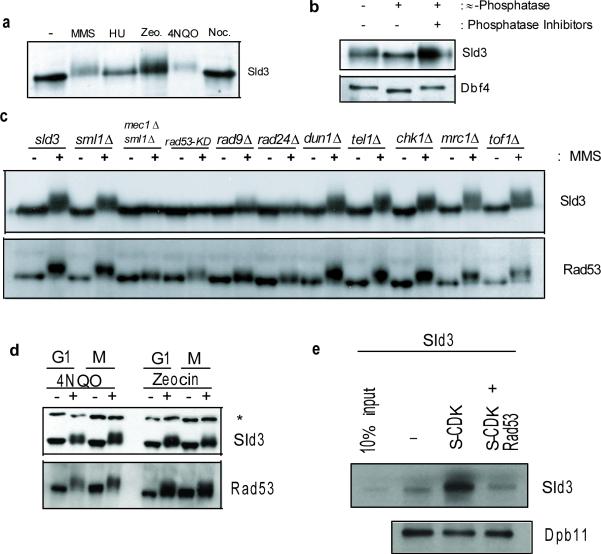
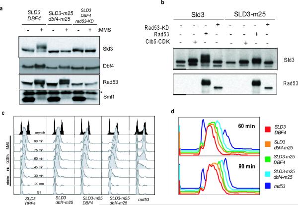
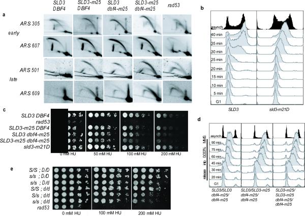
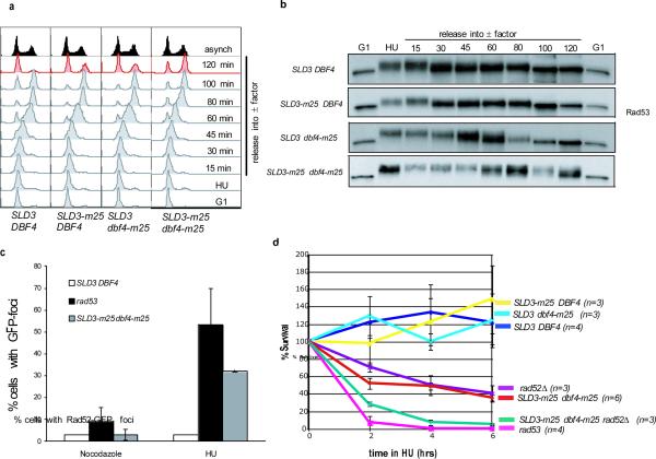
Origins of replication are activated throughout the S phase of the cell cycle such that some origins fire early and others fire late to ensure that each chromosome is completely replicated in a timely fashion. However, in response to DNA damage or replication fork stalling, eukaryotic cells block activation of unfired origins. Human cells derived from patients with ataxia telangiectasia are deficient in this process due to the lack of a functional ataxia telangiectasia mutated (ATM) kinase and elicit radioresistant DNA synthesis after γ-irradiation(2). This effect is conserved in budding yeast, as yeast cells lacking the related kinase Mec1 (ATM and Rad3-related (ATR in humans)) also fail to inhibit DNA synthesis in the presence of DNA damage. This intra-S-phase checkpoint actively regulates DNA synthesis by inhibiting the firing of late replicating origins, and this inhibition requires both Mec1 and the downstream checkpoint kinase Rad53 (Chk2 in humans). However, the Rad53 substrate(s) whose phosphorylation is required to mediate this function has remained unknown. Here we show that the replication initiation protein Sld3 is phosphorylated by Rad53, and that this phosphorylation, along with phosphorylation of the Cdc7 kinase regulatory subunit Dbf4, blocks late origin firing in Saccharomyces cerevisiae. Upon exposure to DNA-damaging agents, cells expressing non-phosphorylatable alleles of SLD3 and DBF4 (SLD3-m25 and dbf4-m25, respectively) proceed through the S phase faster than wild-type cells by inappropriately firing late origins of replication. SLD3-m25 dbf4-m25 cells grow poorly in the presence of the replication inhibitor hydroxyurea and accumulate multiple Rad52 foci. Moreover, SLD3-m25 dbf4-m25 cells are delayed in recovering from transient blocks to replication and subsequently arrest at the DNA damage checkpoint. These data indicate that the intra-S-phase checkpoint functions to block late origin firing in adverse conditions to prevent genomic instability and maximize cell survival.


Figures

Figure 1
Figure 1. Rad53-dependent phosphorylation of Sld3
a, Immunoblotting of asynchronous cells expressing Sld3-3Flag in the absence (−) or presence of MMS (alkylating agent-0.05%), HU (ribonucleoside reductase inhibitor-200 mM), zeocin (radiation mimetic-200 μg/mL), 4NQO (UV-mimetic-2 μg/mL), or nocodazole (10 μg/mL) for 90 minutes. b, Phosphatase assays of Sld3-3Flag and Dbf4-TAP purified from MMS-treated cells. c, Immunoblot of checkpoint deletion mutants expressing Sld3-9myc. Samples were taken from cells arrested in G1 with α-factor and released in the presence or absence of MMS for 180 minutes. d, Immunoblot of cells expressing Sld3-9myc arrested in G1 with α-factor or metaphase with nocodazole for 120 minutes and then treated with either 4NQO or Zeocin, as in a, for 60 minutes. * Denotes a background band. e, In vitro binding assay. Recombinant Sld3-3Flag mock phosphorylated (−), Clb5-TAP CDK phosphorylated or Clb5-TAP CDK phosphorylated followed by Rad53 phosphorylation. 10% of the untreated Sld3-3Flag is shown as input. Immobilized Dpb11-TAP was used to pull down the differentially phosphorylated Sld3 species.
Figure 2
Figure 2. SLD3-m25 and dbf4-m25 cells are intra-S-phase checkpoint defective
a, Immunoblots from asynchronous cells grown in the presence or absence of 0.05% MMS for 90 minutes. rad53-KD (K227A) is a hypomorphic allele that is checkpoint-defective, but able to perform the essential function rescued by Sml1 deletion. b, Immunoblots of an in vitro kinase assay on recombinant full-length Sld3 or Sld3-m25 substrates treated with yeast clb5-TAPCDK, recombinant Rad53 or Rad53-KD (K227A) c, Flow cytometry of wild-type, SLD3-m25, dbf4-m25, SLD3-m25 dbf4-m25, or rad53Δ cells synchronized in G1 with α-factor and released into 0.033% MMS at 30°C. d, The 60 and 90 minute profiles from c, displayed together.
Figure 3
Figure 3. Inappropriate firing of late origins in the presence of replication inhibitors
a, Two-dimensional gel analysis of replication intermediates. Wild-type, SLD3-m25, dbf4-m25, SLD3-m25 dbf4-m25 or rad53Δ cells were synchronized with α-factor and released into 200 mM HU. Cells were collected at 30, 40, 50, 60 and 70 minutes into the HU release and pooled. 20 μg aliquots of DNA were analyzed by two-dimensional electrophoresis as previously described. early origins; ARS305 and ARS607. late origins; ARS501 and ARS609. b, Flow cytometry of cells expressing SLD3-3Flag or a phosphorylation-mimetic allele sld3-m21D-3Flag. Cells were arrested in G1 with α-factor and released into media without α-factor at 30°C. c, 5-fold serial dilutions of wild-type or mutant strains grown on 0 mM, 50 mM, 100 mM, or 200 mM HU. d, Flow cytometry of diploid MAT a/a cells treated as in 2c, from SLD3/SLD3 dbf4-m25/dbf4-m25, SLD3/SLD3-m25 dbf4-m25/dbf4-m25, and SLD3-m25/SLD3-m25 dbf4-m25/dbf4-m25 cells. e, 5-fold serial dilutions, as in c, of diploid strains to assess dominance. Capital S and D represent wild-type SLD3 and DBF4, whereas lowercase s and d represent SLD3-m25 and dbf4-m25 respectively.
Figure 4
Figure 4. Inappropriate firing of late origins elicits DNA damage
a, Flow cytometry of wild-type or mutant strains synchronized in G1 with α-factor, released into a 200 mM HU block for 2 hours, then released into medium with α-factor. b, Immunoblots of samples from a, taken at indicated time points and probed for Rad53. c, Quantification of cells with one or more Rad52-GFP foci after incubating for 90 minutes in the presence of nocodazole or 200 mM HU. Error bars represent s.e.m.; n = 3. d, Survival assay of strains synchronized in G1 and released into 200 mM HU for 2, 4 or 6 hours. Error bars represent s.e.m; n is indicated on graph.

Similar articles

Cited by

References

    1. Painter RB, Young BR. Radiosensitivity in ataxia-telangiectasia: a new explanation. Proc Natl Acad Sci U S A. 1980;77:7315–7317. - PMC - PubMed
    1. Young BR, Painter RB. Radioresistant DNA synthesis and human genetic diseases. Hum Genet. 1989;82:113–117. - PubMed
    1. Larner JM, Lee H, Hamlin JL. Radiation effects on DNA synthesis in a defined chromosomal replicon. Mol Cell Biol. 1994;14:1901–1908. - PMC - PubMed
    1. Paulovich AG, Hartwell LH. A checkpoint regulates the rate of progression through S phase in S. cerevisiae in response to DNA damage. Cell. 1995;82:841–847. - PubMed
    1. Santocanale C, Diffley JF. A Mec1- and Rad53-dependent checkpoint controls late-firing origins of DNA replication. Nature. 1998;395:615–618. - PubMed

Publication types

MeSH terms

Substances