

Review
doi: 10.1038/nrc2931.
Throwing the cancer switch: reciprocal roles of polycomb and trithorax proteins
Affiliations
- PMID: 20865010
- PMCID: PMC4068012
- DOI: 10.1038/nrc2931
Item in Clipboard
Review
Throwing the cancer switch: reciprocal roles of polycomb and trithorax proteins
Nat Rev Cancer.
2010 Oct.
Abstract
The discovery that cancer can be governed above and beyond the level of our DNA presents a new era for designing therapies that reverse the epigenetic state of a tumour cell. Understanding how altered chromatin dynamics leads to malignancy is essential for controlling tumour cells while sparing normal cells. Polycomb and trithorax group proteins are evolutionarily conserved and maintain chromatin in the 'off' or 'on' states, thereby preventing or promoting gene expression, respectively. Recent work highlights the dynamic interplay between these opposing classes of proteins, providing new avenues for understanding how these epigenetic regulators function in tumorigenesis.
Figures
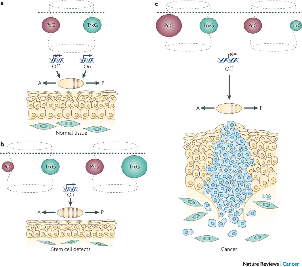
The hourglass represents developmental time, with the establishment and maintenance of chromatin states being depicted by the upper and lower portions of the hourglass, respectively. The hourglass is ‘flexible,’ as alterations in chromatin structure affects gene expression in a highly dynamic manner. a. The chromatin state established by transcription factors during development (above dotted line) is maintained by an evolutionarily conserved mechanism involving a balance between PcG (pink) and TrxG (green) proteins (below dotted line). PcG and TrxG proteins regulate chromatin to evoke transcriptionally inactive (“off”) or active (“on”) states, respectively, thereby maintaining cellular memory. This PcG-TrxG-modulated cellular memory system maintains the identity of body segments along the anterior (A) - posterior (P) axis of the embryo, and regulates tissue homeostasis in the adult. b. Perturbation of cellular memory due to compromised PcG or excessive TrxG leads to inappropriate segment identity and defects in stem cell renewal. c. Perturbation of cellular memory due to excessive PcG or compromised TrxG leads inappropriate segment identity and cancer.
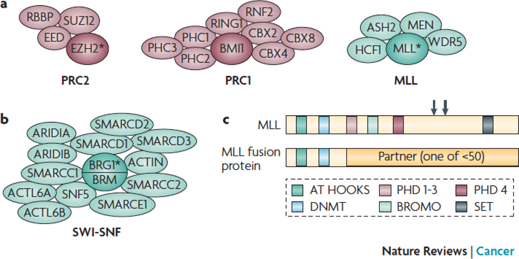
a. PcG complexes PRC2 and PRC1 (left) and the TrxG MLL complex (right) directly methylate histone tails via the HMT activity of the subunit depicted by an asterisk. b. Some TrxG complexes such as SWI-SNF mobilize nucleosomes via the ATPase activity of the subunit depicted by an asterisk. c. Other TrxG proteins such as MLL directly methylate histone tails via the HMT activity encoded by the SET domain (upper diagram). The common breakpoint region (large arrow) and TASPASE cleave sites (small arrows) are shown. Translocations involving MLL (lower diagram) are found in human cancer. These fusions typically retain the MLL N-terminal region, and are devoid of HMT activity.
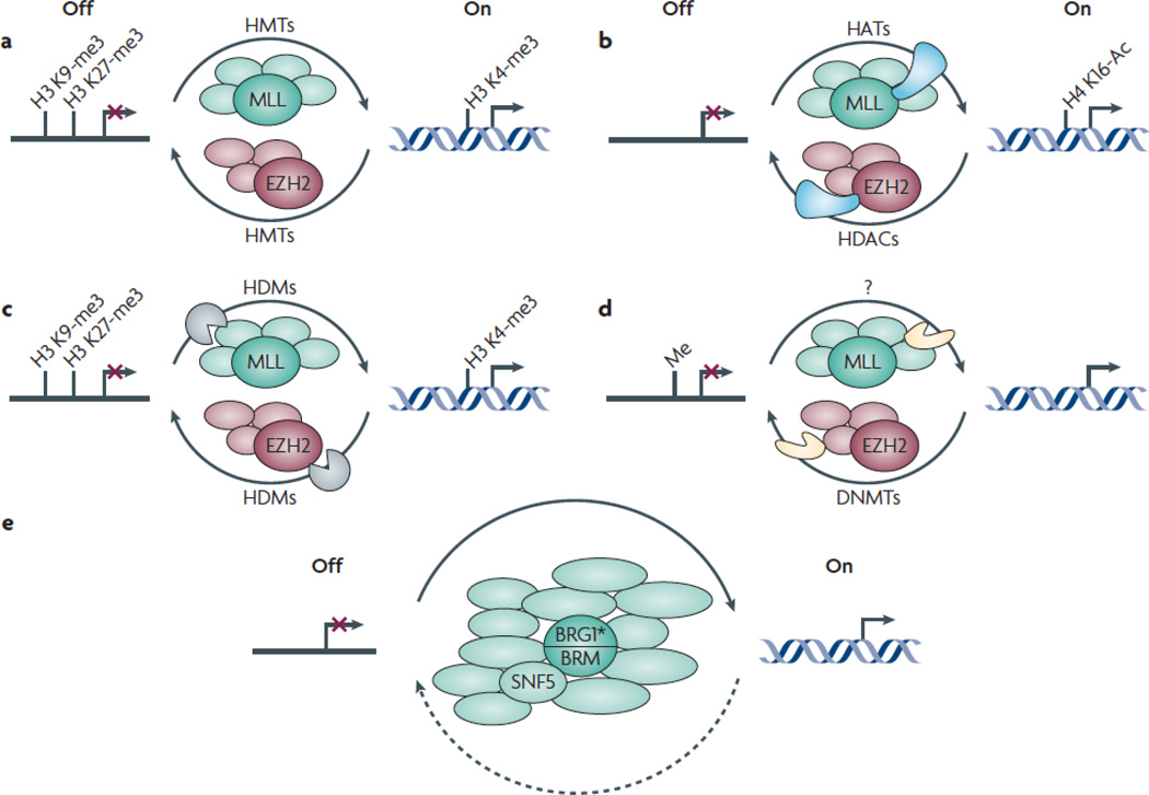
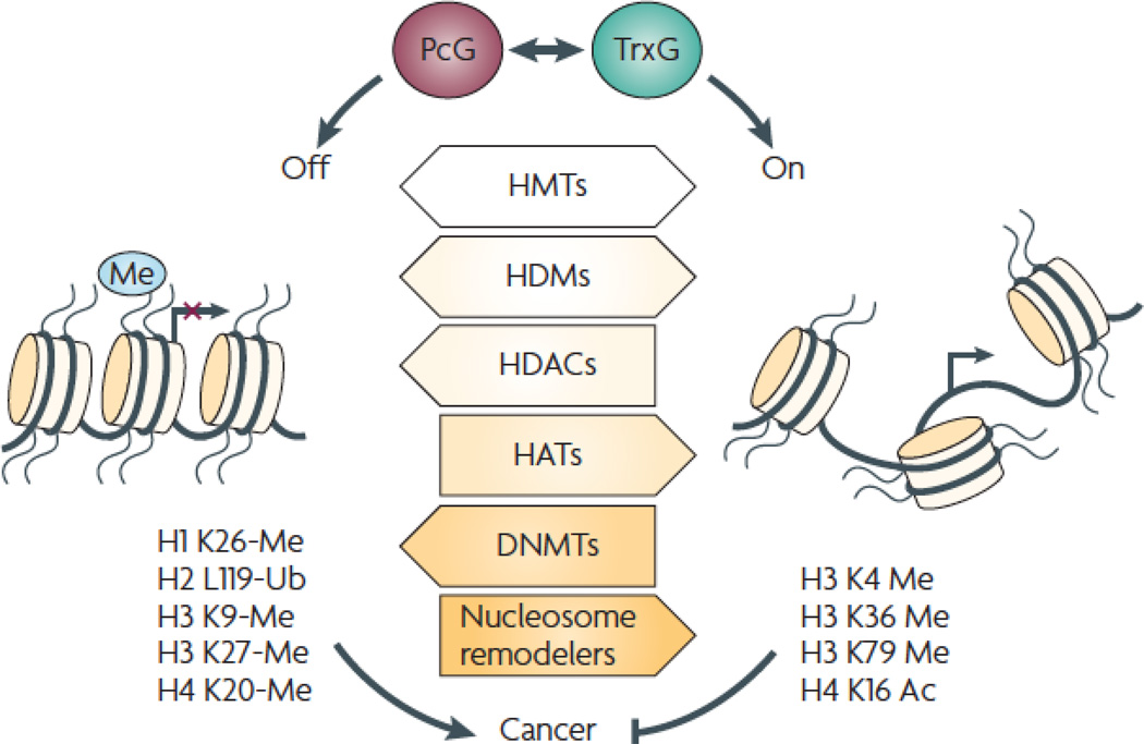
PcG and TrxG-mediated regulation involves multiple enzymatic activities. The mechanisms depicted below are not mutually exclusive. a. PcG and TrxG proteins directly methylate specific histone residues. The PcG and TrxG proteins EZH2 (pink) and MLL (green) are HMTs that establish repressive and activating histone marks, respectively. b. PcG and TrxG complexes recruit enzymes that modulate acetylation of histone residues (blue). Whereas PcG complexes can associate with HDACs that deacetylate histone residues, TrxG complexes can associate with HATs that acetylate histone residues. c. PcG and TrxG complexes recruit enzymes (brown) that remove specific methyl marks from histones. Whereas PcG complexes contain HDMs that remove activating marks, TrxG complexes contain HDMs that remove repressive histone marks. d. PcG and TrxG complexes modulate enzymes (orange) that methylate DNA. Whereas PcG complexes recruit DNMTs that methylate and thereby transcriptionally silence specific genes, TrxG complexes evict DNMTs. e. TrxG proteins remodel nucleosomes. The TrxG proteins BRG1 and BRM are ATP-dependent nucleosome remodelers (green).
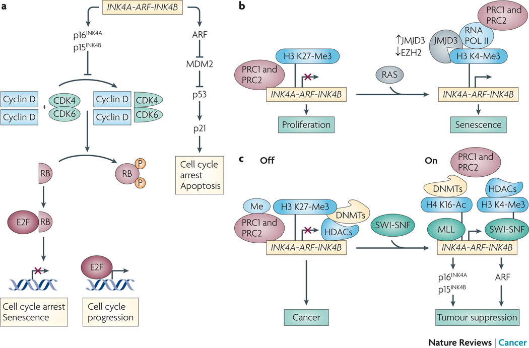
a. The INK4/ARF locus (Ink4/Arf locus in mouse) encodes three tumor suppressors, p15INK4b, ARF (also called p19Arf in mouse) and p16INK4a. p16INK4a induces cell cycle arrest/senescence by inhibiting E2F-mediated transcription of target genes necessary for cell cycle progression. ARF induces cell cycle arrest/apoptosis by facilitating p53 activity via inhibition of MDM2-mediated degradation of p53. p16INK4a/RB- and ARF/p53-mediated tumor suppressive pathways play a nodal role in tumorigenesis. PcG and TrxG proteins function as repressors and activators of the INK4/ARF locus, respectively. b. Normal proliferating cells keep senescence in check by repressing the INK4a-ARFINK4b locus. PcG complexes PRC1 and PRC2 bind INK4a-ARF-INK4b under proliferating conditions, thereby inhibiting senescence. Expression of activated RAS results in recruitment of JMJD3 and pol II, and eviction of PRC proteins. c. TrxG-mediated chromatin dynamics can override PcG-mediated repression in cancer cells. PcG proteins PRC1 and PRC2 are localized at the INK4/ARF locus. This coincides with the recruitment of HDACs, DNMTs, and DNA methylation at the repressed locus in cancer cells. SWI/SNF recruitment to INK4/ARF causes displacement of PcG proteins, DNMTs, and HDACs. MLL is recruited, and the locus is marked with active chromatin marks such as H4K16Ac and H3K4me3, as well as decreased DNA methylation. This coincides with transcriptional activation and activation of p16INK4a-RB- and ARF-p53-mediated tumor suppression. Based on findings from .
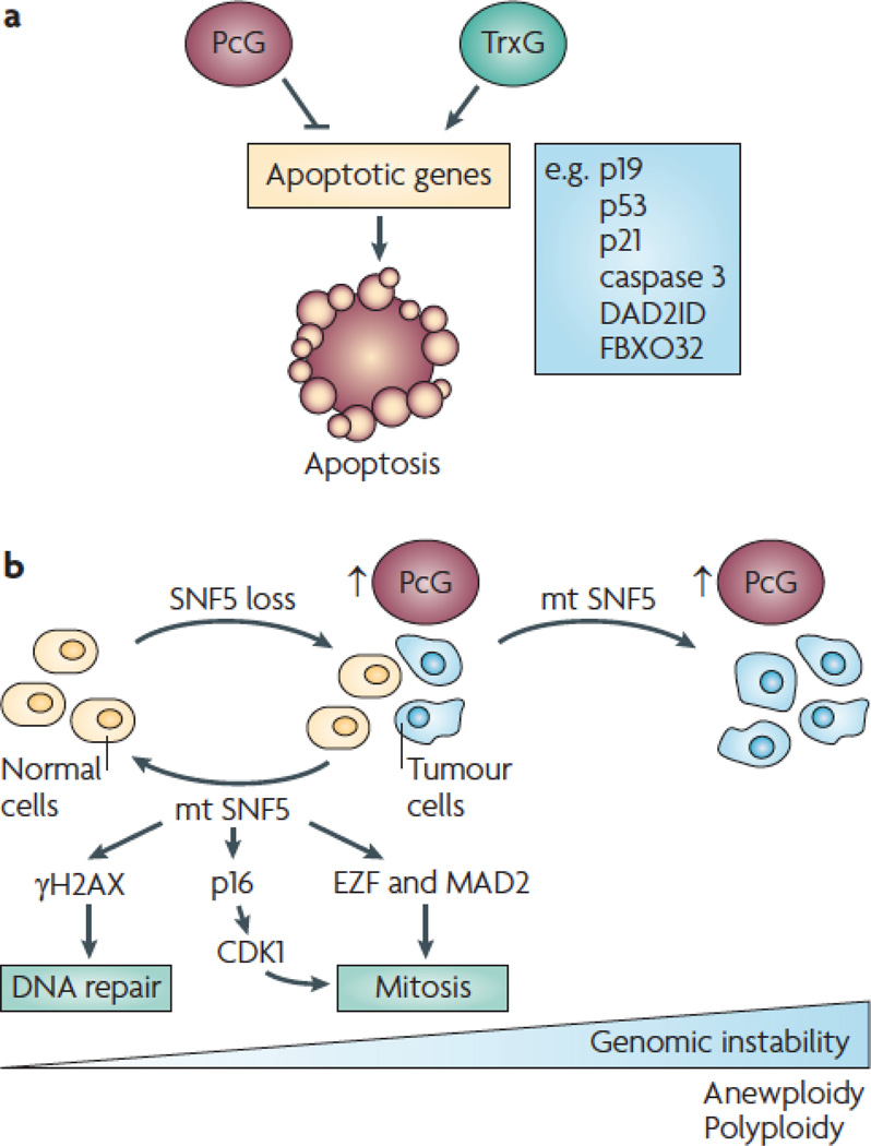
a. PcG and TrxG proteins repress- and activate expression of genes required for apoptosis. b. Loss of the TrxG protein SNF5 leads to genomic instability and cancer. Re-expression of wild type SNF5 in reestablishes a diploid population of cells, whereas expression of tumor-derived SNF5 exacerbates the polyploidy and aneuploid population of cells.
References
-
- Wang JCa, D JE. Cancer stem cells: Lessons from leukemia. Trends Cell Biol. 2005;15:494–501. - PubMed
-
- Clarke MF, et al. Cancer stem cells--perspectives on current status and future directions: AACR Workshop on cancer stem cells. Cancer Res. 2006;66:9339–9344. - PubMed
-
- Campbell LL, Polyak K. Breast tumor heterogeneity: cancer stem cells or clonal evolution? Cell Cycle. 2007;6:2332–2338. - PubMed
-
- Pardal R, Clarke MF, Morrison SJ. Applying the principles of stem-cell biology to cancer. Nature Reviews Cancer. 2003;3:895–902. - PubMed
-
- Hill RP. Identifying cancer stem cells in solid tumors: case not proven. Cancer Res. 2006;66:1891–1895. discussion 1890. - PubMed
Publication types
MeSH terms
Substances
Grants and funding
LinkOut - more resources
Full Text Sources
Other Literature Sources
Molecular Biology Databases
