Review
. 2010 Oct;10(10):707-19.
doi: 10.1038/nrc2888.

Kaposi's sarcoma and its associated herpesvirus

Affiliations
Review

Kaposi's sarcoma and its associated herpesvirus

Enrique A Mesri et al. Nat Rev Cancer. 2010 Oct.

Abstract

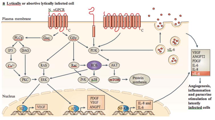
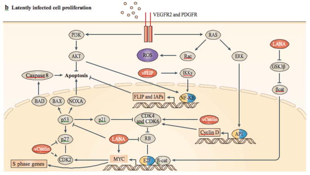
Kaposi's sarcoma (KS) is the most common cancer in HIV-infected untreated individuals. Kaposi's sarcoma-associated herpesvirus (KSHV; also known as human herpesvirus 8 (HHV8)) is the infectious cause of this neoplasm. In this Review we describe the epidemiology of KS and KSHV, and the insights into the remarkable mechanisms through which KSHV can induce KS that have been gained in the past 16 years. KSHV latent transcripts, such as latency-associated nuclear antigen (LANA), viral cyclin, viral FLIP and viral-encoded microRNAs, drive cell proliferation and prevent apoptosis, whereas KSHV lytic proteins, such as viral G protein-coupled receptor, K1 and virally encoded cytokines (viral interleukin-6 and viral chemokines) further contribute to the unique angioproliferative and inflammatory KS lesions through a mechanism called paracrine neoplasia.


Figures

FIGURE 1
FIGURE 1. Geographical prevalence of KS and seroprevalence of KSHV
The standardized incidence of Kaposi’s sarcoma (KS) is depicted for males, and was obtained from the International Agency for Research on Cancer Cancer Incidence in Five Continents publication (see Further information). The rate provided for the United States is an average, but rates in some States (including, California, New York, Georgia and the District of Columbia) can be as high as 6 in some subpopulations. According to Surveillance Epidemiology and End Results (SEER; see Further information) overall rates in the United States among non-Hispanic caucasians is 0.8, among caucasian Hispanics 1.4, and among African Americans is 2.4. In Italy, rates also vary by region, being as low as 0.2 in Umbria but 2.2 in Brescia. Incidences in Africa are taken from the Globocan database (see Further information). b | Seroprevalence rates were compiled from multiple studies. When different rates from the same country are reported, an average was taken. Values represent those in the general population, usually blood donors, and cohorts comprising of HIV-infected individuals were excluded. The seroprevalence of KSHV infection in northern Europe, Asia and the United States is less than 10%, but in most of sub-Saharan Africa, overall seroprevalence is more than 50%. The Mediterranean region has intermediate seroprevalence rates of 10–30%175.
Figure 2
Figure 2. Cellular Heterogeneity in Kaposi’s sarcoma
A biopsy sample from a nodular KS lesion showing numerous spindle cells in the dermis. Immunohistochemical staining shows Kaposi’s sarcoma-associated herpesvirus (KSHV; also known as human herpesvirus 8 (HHV8)) latency-associated nuclear antigen (LANA) in spindle cells lining vascular spaces. The lymphatic marker D2-40 is also localized to vascular spaces. Kaposi’s sarcoma (KS) lesions are composed of various cell types, including vascular (CD34) and lymphatic endothelial cells (D2-40), macrophages (lysozyme), lymphocytes, plasma cells and red blood cells. The inflammatory infiltrate is both inside and outside well-formed or poorly defined vascular spaces. These images highlight the complexity of KS lesions and the presence of virus in only a variable proportion of cells in the lesions, which is consistent with an important role of paracrine angiogenic and inflammatory signals. Magnifications: for hematoxylin and eosin (H&E) x10 and x40; for LANA x40; for CD34, D2-40 and lysozyme x20.
FIGURE 3
FIGURE 3. Proposed mechanism of KSHV-induced sarcoma
a | In lytic or abortive lytic-infected cells, expression of Kaposi’s sarcoma-associated herpesvirus (KSHV; also known as human herpesvirus 8 (HHV8)) early lytic genes (such as viral G protein-coupled receptor (vGPCR), K1, viral interleukin-6 (vIL-6) and K15; shown in red) subvert host signalling pathways, leading to the expression and secretion of angiogenic, inflammatory and proliferative factors (including, vascular endothelial growth factor (VEGF), platelet-derived growth factor-β (PDGFB), angiopoietin 2 (ANGPT2), IL-6 and IL-8). This can occur together with intracrine activity and the secretion of vIL-6. b | Secreted factors stimulate their receptors in latently infected cells through a paracrine mechanism, complementing the autocrine (such as the secretion of cytokines by viral FLICE inhibitory protein (vFLIP)) and direct pro-oncogenic activities of KSHV latent genes, such as vFLIP, vcyclin and latency-associated nuclear antigen (LANA), as well as the KSHV-encoded microRNAs. β-cat, β-catenin; CDK, cyclin-dependent kinase; GSK3β, glycogen synthase kinase 3β; HIF, hypoxia-inducible factor; IAPs, inhibitor of apoptosis proteins; NF-κB, nuclear factor-κB; PKC, protein kinase C; PLC, phospholipase C; ROS, reactive oxygen species.
FIGURE 4
FIGURE 4. Mouse model of KSHV-induced KS
a | Nude mice bearing enhanced green fluorescent protein-expressing tumours that are induced by subcutaneous injection of mouse endothelial cells transfected with Karposi’s sarcoma-associated herpesvirus (KSHV; also known as human herpesvirus 8 (HHV8)) KSHV Bac 36 (mECK36). b | Immunoflurorescence analysis of latency-associated nuclear antigen (LANA; white) and the Kaposi’s sarcoma (KS) marker podoplanin (red) in mECK36 tumours showing punctuated LANA staining that is characteristic of episomal KSHV in cell nucleus (DAPI Blue). c | Activation of paracrine and autocrine endothelial stimulation as shown by gene expression data (heat maps) from AIDS-KS41 and from the mECK36 mouse KS model171. Data are grouped by ligands (left) and receptors (right), which are present in both molecular signatures. Open connectors indicate paracrine stimulation by upregulation in KS of at least one of the receptor–ligand pairs; ligand and receptor closed connectors indicate upregulation of both receptor and ligand with potential for both paracine and autocrine stimulation. ANGPT2, angiopoietin 2; CCL5, chemokine, CC motif, ligand 5; CCR5, chemokine, CC motif receptor 5; CXCL12, chemokine CXC motif, ligand 12; EdnA, Endothelin A; NRP, neuropilin; PDGF, platelet-derived growth factor; TGFβ, transforming growth factor-β; TNF, tumour necrosis factor; VEGF, vascular endothelial growth factor.
Timeline
Timeline
A history of KS KSHV

Similar articles

Cited by

References

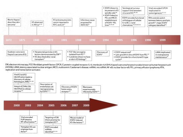
    1. Kaposi M. Idiopathisches multiples Pigmentsarkom der Haut. Arch Dermatol Syph. 1872;4 Original description of KS.
    1. Centers for Disease Control. Kaposi’s sarcoma and Pneumocystis pneumonia among homosexual men- New York City and California. MMWR Morb Mortal Wkly Rep. 1981;30:305–308. - PubMed
    1. Gottlieb GJ, et al. A preliminary communication on extensively disseminated Kaposi’s sarcoma in young homosexual men. Am J Dermatopathol. 1981;3:111–114. - PubMed
    1. Barre-Sinoussi F, et al. Isolation of a T-lymphotropic retrovirus from a patient at risk for acquired immune deficiency syndrome (AIDS) Science. 1983;220:868–871. - PubMed
    1. Gelmann EP, et al. Proviral DNA of a retrovirus, human T-cell leukemia virus, in two patients with AIDS. Science. 1983;220:862–865. - PubMed

Publication types