. 2010 Mar;1(3):239-250.
doi: 10.1177/1947601910368059.

MUC1-C Oncoprotein Interacts Directly with ATM and Promotes the DNA Damage Response to Ionizing Radiation

Affiliations

MUC1-C Oncoprotein Interacts Directly with ATM and Promotes the DNA Damage Response to Ionizing Radiation

Lei Huang et al. Genes Cancer. 2010 Mar.

Abstract

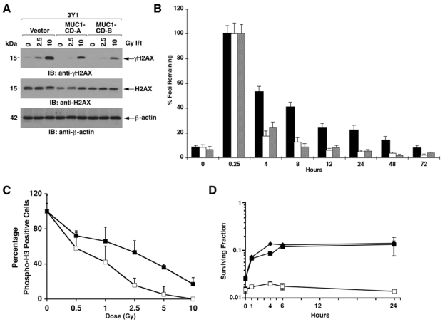
The ataxia-telangiectasia mutated (ATM) kinase is activated in the cellular response to ionizing radiation (IR) and is of importance to the repair of DNA double strand breaks (DSBs). The MUC1 oncoprotein is aberrantly overexpressed in human breast carcinomas. The present work demonstrates that the MUC1 C-terminal subunit (MUC1-C) constitutively interacts with ATM in human breast cancer cells. We show that the MUC1-C cytoplasmic domain binds directly to ATM HEAT repeats. Our results also demonstrate that the MUC1-C cytoplasmic domain binds to the ATM substrate H2AX. The functional significance of these interactions is supported by the finding that MUC1-C promotes removal of IR-induced nuclear γH2AX foci. MUC1-C also protects against IR-induced chromosomal aberrations. In concert with these results, MUC1-C blocks IR-induced death by promoting repair of potentially lethal DNA damage. These findings indicate that the overexpression of MUC1 can protect against IR-induced DNA DSBs and may represent a physiologic response that has been exploited by malignant cells.


Conflict of interest statement

Dr. Kharbanda is an employee of Genus Oncology. Dr. Kufe is a founder of Genus Oncology and a consultant to the company. The other authors declare no potential conflicts of interest.

Figures

Figure 1.
Figure 1.
MUC1-C associates with ataxia-telangiectasia mutated (ATM) kinase. (A) Lysates from ZR-75-1 cells were immunoprecipitated with a control IgG, anti-MUC1-C (left), or anti-ATM (right). The precipitates were immunoblotted with the indicated antibodies. (B) Control IgG and anti-ATM precipitates from MCF-7 cell lysates were immunoblotted with the indicated antibodies. (C) 293 cells were cotransfected with Flag-tagged ATM and MUC1. After 36 h, lysates were immunoprecipitated with a control IgG, anti-MUC1-C (left), or anti-Flag (right). The precipitates were immunoblotted with the indicated antibodies. (D) 293 cells were transfected with green fluorescent protein (GFP) or GFP-MUC1-CD for 36 h. Anti-ATM precipitates were immunoblotted with the indicated antibodies (upper 2 panels). Lysates not subjected to precipitation were immunoblotted with anti-GFP (lower panel).
Figure 2.
Figure 2.
MUC1-CD binds directly to ataxia-telangiectasia mutated (ATM) kinase. (A) Schematic representation of ATM structure and expression of the glutathione S-transferase (GST)–ATM fusion proteins. (B) Lysates from 293 cells expressing green fluorescent protein (GFP)–MUC1-CD were incubated with GST and the indicated GST-ATM fusion proteins. The adsorbates were immunoblotted with anti-GFP. (C) GST and the indicated GST-ATM fusion proteins linked to glutathione beads were incubated with purified His-MUC1-CD. The adsorbates were immunoblotted with anti-MUC1-C. (D) Amino acid sequence of MUC1-CD (upper panel). Lysates from 293 cells expressing GFP or the indicated GFP-MUC1-CD fusion proteins were incubated with GST-ATM(1439-1770). The adsorbates were immunoblotted with anti-GFP. Expression of the GFP proteins was assessed by immunoblot analysis of lysates with anti-GFP (lower panel).
Figure 3.
Figure 3.
MUC1-C associates with the ataxia-telangiectasia mutated (ATM)–H2AX complex. (A) Lysates from 293 cells expressing green fluorescent protein (GFP), GFP-MUC1-CD, GFP-MUC1-CD(C1A), GFP-MUC1-CD(C3A), or GFP-MUC1-CD(C1A/C3A) were incubated with glutathione S-transferase (GST)–ATM(1439-1770). The adsorbates were immunoblotted with anti-GFP (upper panel) and anti-GST (middle panel). Lysates were also immunoblotted with anti-GFP (lower panel). (B) Lysates from 293 cells expressing GFP or the indicated GFP-MUC1-CD fusion proteins were incubated with GST-ATM(1764-2138), GST-ATM(2141-2428), or GST-ATM(2842-3056). Adsorbates were immunoblotted with anti-GFP. Lysates were also immunoblotted with anti-GFP (lower panel). (C) Lysates from 293 cells expressing GFP or the indicated GFP-MUC1-CD fusion proteins were incubated with GST-H2AX. Adsorbates were immunoblotted with anti-GFP (upper panel) and anti-GST (middle panel). Lysates were also immunoblotted with anti-GFP (lower panel). (D) GST and GST-H2AX linked to glutathione beads were incubated with purified His-MUC1-CD. The adsorbates were immunoblotted with anti-MUC1-C (upper panel). Input of GST-H2AX and His-MUC1-CD was determined by immunoblotting with anti-GST and anti-MUC1-C, respectively (lower 2 panels).
Figure 4.
Figure 4.
Silencing MUC1 is associated with increased levels of γH2AX in the response to ionizing radiation (IR). (A) The indicated ZR-75-1 cells were left untreated or treated with 2.5 or 5.0 Gy IR and incubated for 1 h. Lysates were immunoblotted with the indicated antibodies. (B) ZR-75-1/vector and ZR-75-1/MUC1siRNA cells were treated with 10 Gy IR and harvested after incubation for 12 h. Cells were fixed, incubated with PI, and analyzed for cell cycle distribution. (C) ZR-75-1/vector (open squares) and ZR-75-1/MUC1siRNA (solid squares) cells were treated with the indicated doses of IR, incubated for 1 h, and analyzed for reactivity with anti-phospho-H3. The results are expressed as the relative number of phospho-H3-positive cells (mean ± SD of 3 experiments) after IR treatment compared to that obtained with unirradiated cells. (D) ZR-75-1/MUC1siRNA cells were transfected to express an empty vector, Flag-MUC1-CD, or Flag-MUC1-CD(C1A/C3A). The cells were left untreated or treated with 2.5 or 10 Gy IR and incubated for 1 h. Lysates were immunoblotted with the indicated antibodies.
Figure 5.
Figure 5.
MUC1 promotes removal of ionizing radiation (IR)–induced γH2AX foci arrest and attenuates loss of survival. (A) The indicated ZR-75-1 cells were left untreated or treated with 5 Gy IR and incubated for 1 h before staining for γH2AX (red). Nuclei were stained with 4′-6-diamidino-2-phenylindole (DAPI; blue) and the images merged. (B) The indicated ZR-75-1 cells were treated with 10 Gy IR and harvested at the indicated times. Lysates were immunoblotted with the indicated antibodies. (C) ZR-75-1/vector (solid bars) and ZR-75-1/MUC1siRNA (open bars) cells were treated with 1 Gy IR, incubated for the indicated times, and stained with anti-γH2AX. γH2AX foci were counted for at least 30 cells at each time point. The results are expressed as the relative number (mean ± SD) of γH2AX foci/cell remaining compared to that obtained at 0.25 h. (D) ZR-75-1/vector (open squares), ZR-75-1/MUC1siRNA-A (closed squares), and ZR-75-1/MUCsiRNA-B (closed triangles) were treated with the indicated IR doses. After 24 h, the cells were replated and cultured for 2 to 3 weeks. The colonies were stained with crystal violet and counted manually. The results are expressed as the colony number (mean ± SD of 3 determinations) as compared to that for nonirradiated control cells. Plating efficiency for the ZR-75-1/vector, ZR-75-1/MUC1siRNA-A, and ZR-75-1/MUC1siRNA-B cells was 0.11, 0.15 and 0.13, respectively.
Figure 6.
Figure 6.
MUC1-CD is sufficient for promoting γH2AX removal. (A) The indicated 3Y1 cells were left untreated or treated with 2.5 or 10 Gy ionizing radiation (IR) and incubated for 1 h. Lysates were immunoblotted with the indicated antibodies. (B) 3Y1/vector (solid bars) and 3Y1/MUC1-CD-A (open bars) and 3Y1/MUC1-CD-B (shaded bars) cells were treated with 1 Gy IR, incubated for the indicated times, and stained with anti-γH2AX. γH2AX foci were counted for at least 30 cells at each time point. The results are expressed as the relative number (mean ± SD) of γH2AX foci/cell remaining compared to that obtained at 0.25 h. (C) 3Y1/vector (open squares) and 3Y1/MUC1-CD (closed squares) cells were treated with the indicated doses of IR, incubated for 1 h, and analyzed for reactivity with anti-phospho-H3. The results are expressed as the relative number of phospho-H3-positive cells (mean ± SD of 3 experiments) after IR treatment compared to that obtained with unirradiated cells. (D) 3Y1/vector (open squares), 3Y1/MUC1-CD-A (closed squares), and 3Y1/MUC1-CD-B (closed diamonds) cells were treated with 7 Gy IR. The cells were replated at the indicated times. The results are expressed as colony number (mean ± SEM of 3 determinations). SF24/SF0 for the 3Y1/vector, 3Y1/MUC1-CD-A, and 3Y1/MUC1-CD cells was 1.5, 5.4, and 5.7, respectively.

Similar articles

Cited by

References

    1. Lavin M, Kozlov S. ATM activation and DNA damage response. Cell Cycle 2007;6:931-42 - PubMed
    1. Abraham RT. Cell cycle checkpoint signaling through the ATM and ATR kinases. Genes Dev 2001;15:2177-96 - PubMed
    1. Bonner W, Redon C, Dickey J, Nakamura A, Sedelnikova O, Solier S, et al. GammaH2AX and cancer. Nat Rev Cancer 2008;8:957-67 - PMC - PubMed
    1. Burma S, Chen B, Murphy M, Kurimasa A, Chen D. ATM phosphorylates histone H2AX in response to DNA double-strand breaks. J Biol Chem 2001;276:42462-7 - PubMed
    1. Rogakou EP, Pilch DR, Orr AH, Ivanova VS, Bonner WM. DNA double-stranded breaks induce histone H2AX phosphorylation on serine 139. J Biol Chem 1998;273:5858-68 - PubMed

LinkOut - more resources