. 2010 Aug 26;6(8):e1001069.
doi: 10.1371/journal.ppat.1001069.

PKC signaling regulates drug resistance of the fungal pathogen Candida albicans via circuitry comprised of Mkc1, calcineurin, and Hsp90

Affiliations

PKC signaling regulates drug resistance of the fungal pathogen Candida albicans via circuitry comprised of Mkc1, calcineurin, and Hsp90

Shantelle L LaFayette et al. PLoS Pathog. .

Abstract

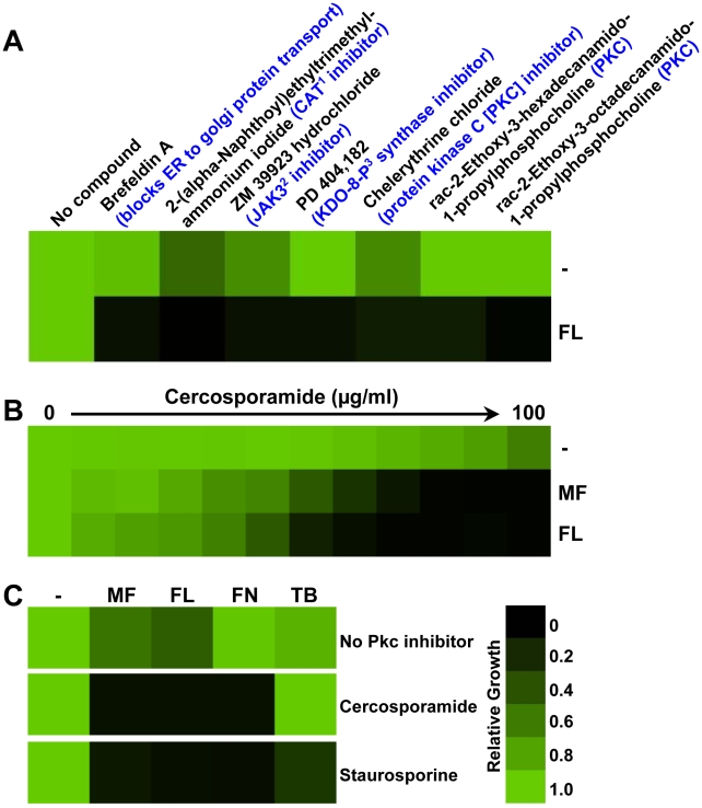
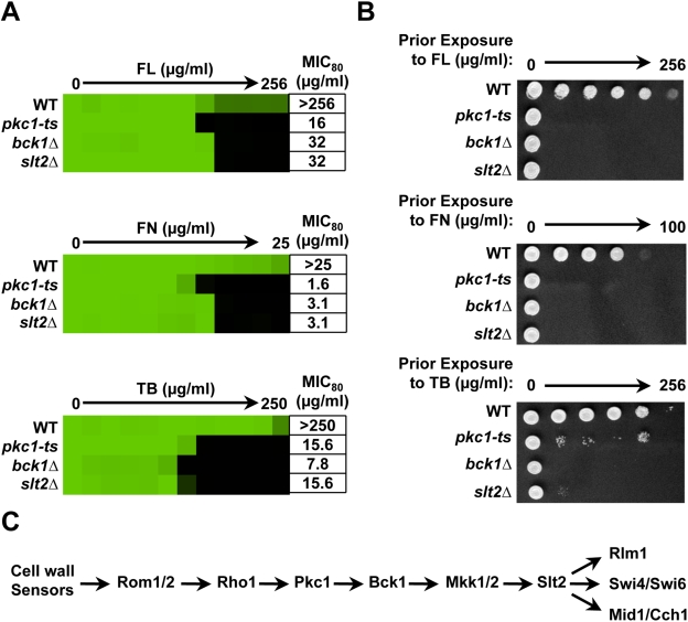
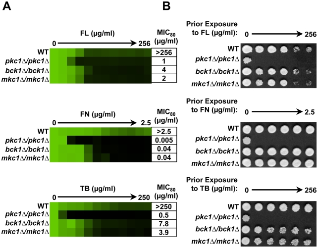
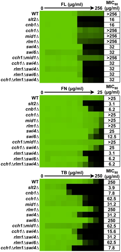
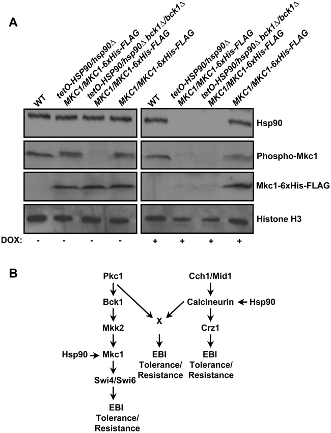
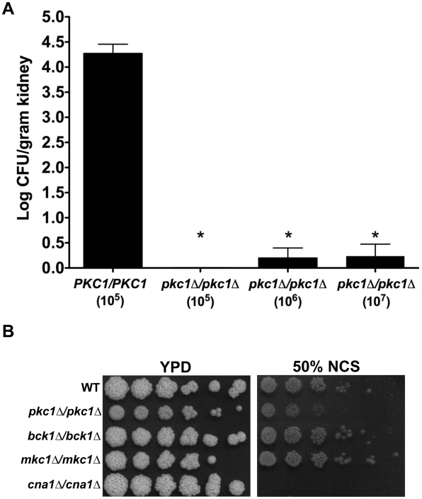
Fungal pathogens exploit diverse mechanisms to survive exposure to antifungal drugs. This poses concern given the limited number of clinically useful antifungals and the growing population of immunocompromised individuals vulnerable to life-threatening fungal infection. To identify molecules that abrogate resistance to the most widely deployed class of antifungals, the azoles, we conducted a screen of 1,280 pharmacologically active compounds. Three out of seven hits that abolished azole resistance of a resistant mutant of the model yeast Saccharomyces cerevisiae and a clinical isolate of the leading human fungal pathogen Candida albicans were inhibitors of protein kinase C (PKC), which regulates cell wall integrity during growth, morphogenesis, and response to cell wall stress. Pharmacological or genetic impairment of Pkc1 conferred hypersensitivity to multiple drugs that target synthesis of the key cell membrane sterol ergosterol, including azoles, allylamines, and morpholines. Pkc1 enabled survival of cell membrane stress at least in part via the mitogen activated protein kinase (MAPK) cascade in both species, though through distinct downstream effectors. Strikingly, inhibition of Pkc1 phenocopied inhibition of the molecular chaperone Hsp90 or its client protein calcineurin. PKC signaling was required for calcineurin activation in response to drug exposure in S. cerevisiae. In contrast, Pkc1 and calcineurin independently regulate drug resistance via a common target in C. albicans. We identified an additional level of regulatory control in the C. albicans circuitry linking PKC signaling, Hsp90, and calcineurin as genetic reduction of Hsp90 led to depletion of the terminal MAPK, Mkc1. Deletion of C. albicans PKC1 rendered fungistatic ergosterol biosynthesis inhibitors fungicidal and attenuated virulence in a murine model of systemic candidiasis. This work establishes a new role for PKC signaling in drug resistance, novel circuitry through which Hsp90 regulates drug resistance, and that targeting stress response signaling provides a promising strategy for treating life-threatening fungal infections.


Conflict of interest statement

The authors have declared that no competing interests exist.

Figures

Figure 1
Figure 1. Pharmacological inhibition of PKC signaling enhances the efficacy of antifungal drugs targeting the cell membrane.
(A) A drug screen identifies compounds that abrogate fluconazole (FL) resistance of a Candida albicans clinical isolate (CaCi-2). Seven compounds from the LOPAC1280 Navigator library had little toxicity on their own but enhanced the efficacy of FL against CaCi-2 when tested at 12.5 µM in RPMI medium with 2% glucose in the presence or absence of 8 µg/ml FL. Growth was measured by absorbance at 600 nm after 48 hours at 30°C. Optical densities were averaged for duplicate measurements and normalized relative to the no compound control (-) or FL-only control. Data was quantitatively displayed with colour using Treeview (see colour bar). The target or mode of action of each compound is indicated in blue. 1CAT = choline acetyltransferase; 2JAK3 = Janus kinase family protein; and 3KDO-8-P = 3-deoxy-D-manno-2-octulosonate-8-phosphate. (B) Pharmacological inhibition of Pkc1 with cercosporamide abrogates azole resistance and reduces echinocandin tolerance of CaCi-2 in an MIC assay. Assays were done in yeast peptone dextrose (YPD) with a fixed concentration of 2 µg/ml micafungin (MF) or 8 µg/ml FL, as indicated. Data was analyzed after 48 hours at 30°C as in part A. (C) Pkc1 inhibitors confer increased sensitivity to other ergosterol biosynthesis inhibitors. A fixed concentration of CaCi-2 cells was incubated in YPD with no antifungal (-), 2 µg/ml MF, 8 µg/ml FL, 2.5 µg/ml fenpropimorph (FN), or 2 µg/ml terbinafine (TB) and with the PKC inhibitors cercosporamide (100 µg/ml) or staurosporine (37.5 ng/ml), as indicated. Data was analyzed after 48 hours at 30°C as in part A.
Figure 2
Figure 2. Pkc1 enables basal tolerance to ergosterol biosynthesis inhibitors via the MAPK cascade in Saccharomyces cerevisiae.
(A) Drug tolerance of a wild-type (WT) strain (BY4741), a derivative (pkc1-ts) with a temperature sensitive PKC1 allele, and derivatives with deletions of BCK1 and SLT2 are compared in MIC assays. Assays were performed in synthetic defined (SD) medium at 35°C. Data was analyzed after 48 hours as in Figure 1A. The minimum drug concentration that inhibits growth by 80% relative to the drug-free growth control (MIC80) is indicated for each strain. (B) Genetic compromise of Pkc1 creates a fungicidal combination with ergosterol biosynthesis inhibitors. MIC assays with two-fold dilutions of fluconazole (FL), fenpropimorph (FN), and terbinafine (TB) were performed in SD and incubated for 48 hours at 35°C. Cells from the MIC assays were spotted onto YPD medium and incubated at 30°C for 48 hours before plates were photographed. (C) Schematic of the S. cerevisiae Pkc1 cell wall integrity pathway.
Figure 3
Figure 3. Pkc1 enables basal tolerance to ergosterol biosynthesis inhibitors in part via the MAPK cascade in Candida albicans.
(A) Deletion of PKC1, BCK1 or MKC1 reduces tolerance to fluconazole (FL), fenpropimorph (FN), and terbinafine (TB) in MIC assays. Assays were performed in YPD medium at 35°C with strains derived from the WT SN95. Data was analyzed after 72 hours growth as in Figure 1A. The minimum drug concentration that inhibits growth by 80% relative to the drug-free growth control (MIC80) is indicated for each strain. (B) Deletion of PKC1, but not MAPK components, creates a fungicidal combination with the ergosterol biosynthesis inhibitors in C. albicans. MIC assays with four-fold dilutions of FL, FN, and TB were performed in YPD and incubated for 48 hours at 35°C. Cells from the MIC assays were spotted onto YPD medium and incubated at 30°C for 48 hours before plates were photographed.
Figure 4
Figure 4. Distinct downstream effectors are important for tolerance of S. cerevisiae to different ergosterol biosynthesis inhibitors.
To dissect the role of downstream effectors of Slt2 in tolerance to ergosterol biosynthesis inhibitors, we tested the impact of their deletion individually and in combination on drug susceptibility in an MIC assay. Data was analyzed after 72 hours at 35°C in SD medium as in Figure 1A. The minimum drug concentration that inhibits growth by 80% relative to the drug-free growth control (MIC80) is indicated for each strain.
Figure 5
Figure 5. Swi4 and Cch1-Mid1 play critical roles in ergosterol biosynthesis inhibitor tolerance of C. albicans.
Deletion of SWI4 or components of the Cch1-Mid1 channel confer increased sensitivity to the ergosterol biosynthesis inhibitors in a MIC assay. Deletion of Rlm1 had no impact on drug sensitivity. A strain lacking the catalytic subunit of calcineurin (Cna1) is included for reference. Data was analyzed after 48 hours in YPD at 35°C as in Figure 1A. The minimum drug concentration that inhibits growth by 80% relative to the drug-free growth control (MIC80) is indicated for each strain.
Figure 6
Figure 6. Compromising PKC-MAPK signaling blocks calcineurin activation in response to ergosterol biosynthesis inhibitors in S. cerevisiae.
(A) Genetically compromising PKC-MAPK signaling by deleting SLT2 blocks calcineurin activation monitored with a 4XCDRE-lacZ reporter. β-galactosidase activity was measured after incubation in SD medium for 24 hours without any antifungal (U) or in the presence of ergosterol biosynthesis inhibitors at the following concentrations: 16 µg/mL fluconazole (FL), 1 µg/mL fenpropimorph (FN), or 25 µg/mL terbinafine (TB). While the WT strain exhibited increased β-galactosidase activity in response to ergosterol biosynthesis inhibitors, deletion of SLT2 or CNB1 (which encodes the regulatory subunit of calcineurin) blocked calcineurin activation. Data are means ± SD for triplicate samples and are representative of two independent experiments. (B) Pharmacological inhibition of PKC signaling with staurosporine (STS) blocks calcineurin activation monitored with a 4XCDRE-lacZ reporter. β-galactosidase activity was measured after incubation in SD medium (-) or in SD with 2.5 µg/mL STS. Cells were then treated with 32 µg/mL FL or were left untreated (U). Data are means ± SD for triplicate samples and are representative of two independent experiments. (C) Simplified schematic of how S. cerevisiae Pkc1 regulates responses to ergosterol biosynthesis inhibitors (EBIs) important for basal tolerance and resistance.
Figure 7
Figure 7. PKC signaling and calcineurin independently regulate tolerance to ergosterol biosynthesis inhibitors via a common target in C. albicans.
(A) Deletion of C. albicans PKC1 does not block EBI-induced activation of calcineurin. Transcript levels of two calcineurin-dependent genes, PLC3 and UTR2, were measured by quantitative RT-PCR after growth in rich medium at 35°C for 6 hours without any antifungal (U) or with 16 µg/mL fluconazole (FL), as indicated. Transcripts were normalized to GPD1. Levels are expressed relative to the untreated wild-type samples, which were set to 1. Data are means ± SD for triplicate samples and are representative of two independent experiments. (B) Simultaneous inhibition of calcineurin and Pkc1 signaling does not synergistically decrease FL tolerance of a WT strain (SN95). A fractional inhibitory concentration (FIC) assay was carried out in YPD medium containing a fixed concentration of 0.5 µg/mL FL and gradients of the calcineurin inhibitor cyclosporin A (CsA) and the PKC inhibitor staurosporine (STS). Data was analyzed after growth at 35°C for 48 hours as in Figure 1A. The minimum concentration of STS or CsA that inhibits growth by 80% relative to the FL-only growth control (MIC80) individually or in combination is indicated along with the FIC. (C) FL tolerance of a mutant lacking the catalytic subunit of calcineurin is not sensitive to inhibition of PKC signaling. MIC assays were performed in YPD medium only (-) or YPD with a fixed concentration of 0.5 µg/mL FL. (D) Simplified schematic of how C. albicans Pkc1 regulates responses to ergosterol biosynthesis inhibitors (EBIs) important for basal tolerance and resistance.
Figure 8
Figure 8. Inhibition of PKC signaling phenocopies inhibition of Hsp90 reducing azole resistance.
(A) Fluconazole (FL) resistance of clinical isolates is abrogated by inhibition of Hsp90 or Pkc1. MIC assays were conducted in YPD medium with no inhibitor (-), with the Hsp90 inhibitor geldanamycin (5 µM), or with the Pkc1 inhibitors cercosporamide (12.5 µg/ml) or staurosporine (0.5 µg/ml). Clinical isolates (CaCi) are ordered sequentially with those recovered early in treatment at the top and those recovered late at the bottom; the FL-sensitive strain SC5314 is included as a control. Data was analyzed after growth for 48 hours at 30°C as in Figure 1A. (B) Inhibition of Hsp90 or Pkc1 abrogates FL resistance of both S. cerevisiae and C. albicans erg3 mutants. Inhibition of Hsp90 or Pkc1 has no effect on the FL resistance of a S. cerevisiae strain (PDR1R) that overexpresses multiple drug efflux pumps due to an activating mutation in the transcription factor Pdr1. FL MIC assays were carried out in YPD medium only (-) or in YPD with fixed concentrations of: geldanamycin (Sc: 5 µM; Ca: 0.625 µM), cercosporamide (Sc: 50 µg/ml; Ca: 25 µg/ml), or STS (Sc: 0.625 µg/ml; Ca: 0.3125 µg/ml). Data was analyzed after growth for 48 hours at 30°C as in Figure 1A. The minimum drug concentration that inhibits growth by 80% relative to the no-FL growth control (MIC80) is indicated for each strain.
Figure 9
Figure 9. Hsp90 stabilizes the terminal MAPK Mkc1 in C. albicans.
(A) Genetic reduction of Hsp90 levels results in depletion of Mkc1. In strains where the sole allele of HSP90 is under the control of a tetracycline repressible promoter (tetO), transcription of HSP90 can be repressed by tetracycline or the analog doxycycline (DOX). One allele of MKC1 was C-terminally 6xHis-FLAG tagged for monitoring total levels of Mkc1. The MAPKKK Bck1 was deleted to block phosphorylation of Mkc1. Cells were grown with or without DOX (20 µg/ml) before being treated for 3 hours with 50 µg/ml terbinafine (TB) to elicit phosphorylation of Mkc1. Total protein was resolved by SDS-PAGE and blots were hybridized with α-Hsp90, α-His6 to monitor total Mkc1 levels, α-phospho p44/42 MAPK to monitor dually phosphorylated Mkc1 levels, and α-H3 as a loading control. (B) Simplified schematic of how C. albicans Hsp90 governs responses to ergosterol biosynthesis inhibitors (EBIs) important for basal tolerance and resistance by regulating both Pkc1-MAPK signaling and calcineurin signaling.
Figure 10
Figure 10. Deletion of C. albicans PKC1 attenuates virulence in a murine model by targets distinct from calcineurin.
(A) CD1 mice were infected with an inoculum of the wild-type strain (SN95) of 1×10⁵ CFU or inoculum of the pkc1Δ/pkc1Δ mutant of 1×10⁵ CFU, 1×10⁶ CFU, or 1×10⁷ CFU. Despite the higher innoculum used, deletion of PKC1 resulted in a dramatic reduction of kidney fungal burden. Asterisks indicate P<0.001 (ANOVA, Bonferroni's Multiple Comparison Test). (B) Deletion of PKC1, but not components of the MAPK cascade, results in a modest increase in sensitivity to serum compared to the hypersensitivity of a mutant lacking the catalytic subunit of calcineurin, Cna1. Cells were spotted in fivefold dilutions (from 1×10⁷ cells/ml for pkc1Δ/pkc1Δ; from 1×10⁶ cells/ml for other strains) onto solid YPD medium with 50% new calf serum (NCS), as indicated. Plates were photographed after 72 hours growth at 35°C.

Similar articles

Cited by

References

    1. Bahn YS, Xue C, Idnurm A, Rutherford JC, Heitman J, et al. Sensing the environment: lessons from fungi. Nat Rev Microbiol. 2007;5:57–69. - PubMed
    1. Cowen LE, Steinbach WJ. Stress, drugs, and evolution: the role of cellular signaling in fungal drug resistance. Eukaryot Cell. 2008;7:747–764. - PMC - PubMed
    1. Camilli A, Bassler BL. Bacterial small-molecule signaling pathways. Science. 2006;311:1113–1116. - PMC - PubMed
    1. Hogan DA. Talking to themselves: autoregulation and quorum sensing in fungi. Eukaryot Cell. 2006;5:613–619. - PMC - PubMed
    1. Keller NP, Turner G, Bennett JW. Fungal secondary metabolism - from biochemistry to genomics. Nat Rev Microbiol. 2005;3:937–947. - PubMed

Publication types

MeSH terms