Controlled Clinical Trial
. 2010 Dec;53(12):2646-55.
doi: 10.1007/s00125-010-1910-x. Epub 2010 Sep 25.

Effect of sulodexide on endothelial glycocalyx and vascular permeability in patients with type 2 diabetes mellitus

Affiliations
Controlled Clinical Trial

Effect of sulodexide on endothelial glycocalyx and vascular permeability in patients with type 2 diabetes mellitus

L N Broekhuizen et al. Diabetologia. 2010 Dec.

Abstract

Aims/hypothesis: Endothelial glycocalyx perturbation contributes to increased vascular permeability. In the present study we set out to evaluate whether: (1) glycocalyx is perturbed in individuals with type 2 diabetes mellitus, and (2) oral glycocalyx precursor treatment improves glycocalyx properties.

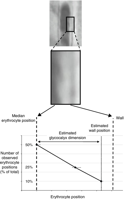
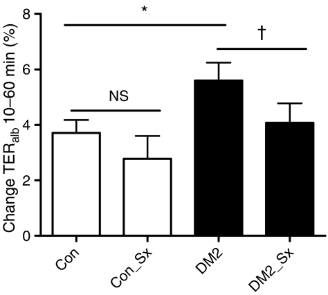
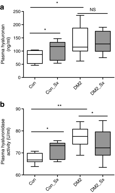
Methods: Male participants with type 2 diabetes (n = 10) and controls (n = 10) were evaluated before and after 2 months of sulodexide administration (200 mg/day). The glycocalyx dimension was estimated in two different vascular beds using sidestream dark field imaging and combined fluorescein/indocyanine green angiography for sublingual and retinal vessels, respectively. Transcapillary escape rate of albumin (TER(alb)) and hyaluronan catabolism were assessed as measures of vascular permeability.

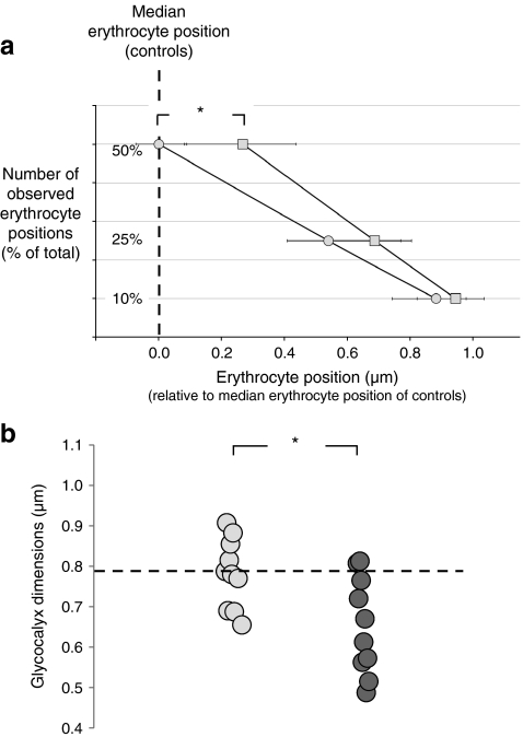
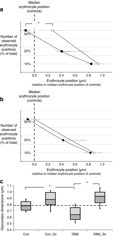
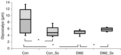
Results: Both sublingual dimensions (0.64 [0.57-0.75] μm vs 0.78 [0.71-0.85] μm, p < 0.05, medians [interquartile range]) and retinal glycocalyx dimensions (5.38 [4.88-6.59] μm vs 8.89 [4.74-11.84] μm, p < 0.05) were reduced in the type 2 diabetes group compared with the controls whereas TER(alb) was increased (5.6 ± 2.3% vs 3.7 ± 1.7% in the controls, p < 0.05). In line with these findings, markers of hyaluronan catabolism were increased with diabetes (hyaluronan 137 ± 29 vs 81 ± 8 ng/ml and hyaluronidase 78 ± 4 vs 67 ± 2 U/ml, both p < 0.05). Sulodexide increased both the sublingual and retinal glycocalyx dimensions in participants with diabetes (to 0.93 [0.83-0.99] μm and to 5.88 [5.33-6.26] μm, respectively, p < 0.05). In line, a trend towards TER(alb) normalisation (to 4.0 ± 2.3%) and decreases in plasma hyaluronidase (to 72 ± 2 U/ml, p < 0.05) were observed in the diabetes group.

Conclusion/interpretation: Type 2 diabetes is associated with glycocalyx perturbation and increased vascular permeability, which are partially restored following sulodexide administration. Further studies are warranted to determine whether long-term treatment with sulodexide has a beneficial effect on cardiovascular risk.

Trial registration: www.trialregister.nl NTR780/ http://isrctn.org ISRCTN82695186

Funding: An unrestricted Novartis Foundation for Cardiovascular Excellence grant (2006) to M. Nieuwdorp/E. S. G. Stroes, Dutch Heart Foundation (grant number 2005T037).


Figures

Fig. 1
Fig. 1
Measurement of sublingual glycocalyx dimension using SDF imaging
Fig. 2
Fig. 2
Endothelial glycocalyx in control and type 2 diabetic participants. a Compared with controls, median erythrocyte width was significantly increased in participants with type 2 diabetes, resulting in a significant movement of erythrocytes towards the vessel wall; *p < 0.05. Circles, controls; squares, type 2 diabetes group. b Glycocalyx dimensions were reduced in the group with type 2 diabetes because of a significant increase in the median erythrocyte width, while both control and diabetic participants had similar values of the 90th percentiles of the erythrocyte column width (identifying the position of the vessel wall), *p < 0.05. Light circles, controls; dark circles, type 2 diabetes group
Fig. 3
Fig. 3
The effect of sulodexide on endothelial glycocalyx in control and diabetic participants. a Following sulodexide treatment, median erythrocyte width decreased significantly in the type 2 diabetes group, resulting in a significant restoration of the distance of erythrocytes from the vessel wall; *p < 0.05. White squares, diabetes group before sulodexide exposure; black squares, diabetes group following sulodexide treatment. b Following sulodexide treatment, median erythrocyte width was not significantly changed in controls, resulting in similar distances of erythrocytes from the vessel. White circles, control group before sulodexide exposure; black circles, diabetes group following sulodexide treatment. c Glycocalyx dimensions at baseline and after 8 weeks of treatment with sulodexide in controls (n = 10; con and Con_Sx, respectively) and diabetic participants (n = 10; DM2 and DM2_Sx, respectively) measured by SDF imaging show sublingual glycocalyx dimensions were significantly reduced in type 2 diabetes, and sulodexide treatment restored microvascular glycocalyx dimensions to control values. Individual capillary glycocalyx dimensions were estimated from the transient widening of the erythrocyte column. Data are presented as whisker plots with median (interquartile range). *p < 0.05
Fig. 4
Fig. 4
The effect of sulodexide on retinal glycocalyx in control and diabetic participants. Glycocalyx dimensions in controls at baseline and after 8 weeks of treatment with sulodexide (n = 6; con and Con_Sx, respectively) and diabetic participants (n = 6; DM2 and DM2_Sx, respectively) measured by FAG/ICG. Individual glycocalyx dimensions were estimated by subtracting the intravascular distribution of two different fluorescent tracers: indocyanine green from fluorescein. Data are presented as whisker plots with median (interquartile range). *p < 0.05
Fig. 5
Fig. 5
The effect of sulodexide on vascular permeability in control and diabetic participants. Percentage change in TERalb at baseline and after 8 weeks of treatment with sulodexide in controls (n = 10; con and Con_Sx) and diabetic participants (n = 10; DM2 and DM2_Sx); p = NS for comparison of controls and controls following sulodexide treatment. Microvascular permeability was determined by the transcapillary escape rate of 125I-labelled albumin (TERalb) between 10 and 60 min after infusion. Data are presented as means ± SD. *p < 0.05, p = 0.08
Fig. 6
Fig. 6
The effect of sulodexide on plasma glycosaminoglycans in control and diabetic participants. a Circulating plasma hyaluronan levels at baseline and after 8 weeks of sulodexide administration in controls (n = 10; con and Con_Sx, respectively) and diabetic participants (n = 10; DM2 and DM2_Sx, respectively). b Hyaluronidase levels at baseline and after 8 weeks of sulodexide administration in controls (n = 10; con and Con_Sx, respectively) and diabetic participants (n = 10; DM2 and DM2_Sx, respectively), p < 0.05 for comparison of controls and controls following sulodexide treatment; p < 0.01 for comparison of controls, controls following sulodexide treatment and diabetes group before sulodexide treatment; p < 0.05 for diabetes group before and after sulodexide treatment. Data are presented as whisker plots with median (interquartile range). *p < 0.05, **p < 0.01

References

    1. Deckert T, Feldt-Rasmussen B, Borch-Johnsen K, Jensen T, Kofoed-Enevoldsen A. Albuminuria reflects widespread vascular damage. The Steno hypothesis. Diabetologia. 1989;32:219–226. doi: 10.1007/BF00285287. - DOI - PubMed
    1. Gerstein HC, Mann JF, Yi Q, et al. Albuminuria and risk of cardiovascular events, death, and heart failure in diabetic and nondiabetic individuals. JAMA. 2001;25(286):421–426. doi: 10.1001/jama.286.4.421. - DOI - PubMed
    1. Morrish NJ, Wang SL, Stevens LK, Fuller JH, Keen H. Mortality and causes of death in the WHO multinational study of vascular disease in diabetes. Diabetologia. 2001;44(Suppl 2):S14–S21. doi: 10.1007/PL00002934. - DOI - PubMed
    1. Brownlee M. Biochemistry and molecular cell biology of diabetic complications. Nature. 2001;414:813–820. doi: 10.1038/414813a. - DOI - PubMed
    1. Nieuwdorp M, van Haeften TW, Gouverneur MC, et al. Loss of endothelial glycocalyx during acute hyperglycaemia coincides with endothelial dysfunction and coagulation activation in vivo. Diabetes. 2006;55:480–486. doi: 10.2337/diabetes.55.02.06.db05-1103. - DOI - PubMed

Publication types

MeSH terms

Associated data