

Improved discrimination of melanotic schwannoma from melanocytic lesions by combined morphological and GNAQ mutational analysis
- PMID: 20865267
- PMCID: PMC2991233
- DOI: 10.1007/s00401-010-0749-z
Improved discrimination of melanotic schwannoma from melanocytic lesions by combined morphological and GNAQ mutational analysis
Abstract
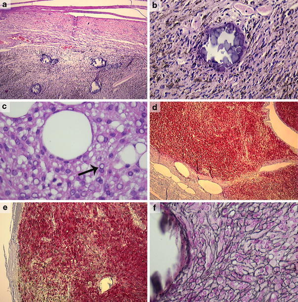
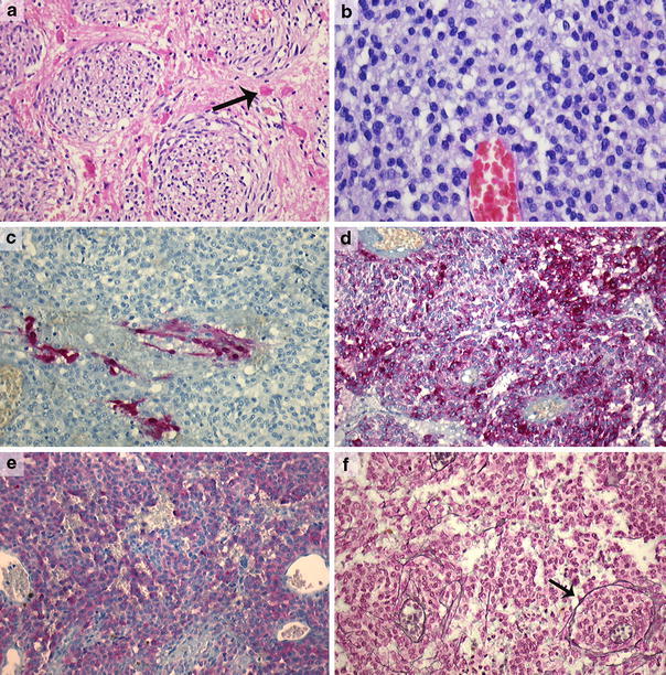
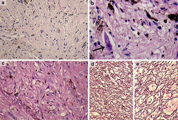
The histological differential diagnosis between melanotic schwannoma, primary leptomeningeal melanocytic lesions and cellular blue nevus can be challenging. Correct diagnosis of melanotic schwannoma is important to select patients who need clinical evaluation for possible association with Carney complex. Recently, we described the presence of activating codon 209 mutations in the GNAQ gene in primary leptomeningeal melanocytic lesions. Identical codon 209 mutations have been described in blue nevi. The aims of the present study were to (1) perform a histological review of a series of lesions (initially) diagnosed as melanotic schwannoma and analyze them for GNAQ mutations, and (2) test the diagnostic value of GNAQ mutational analysis in the differential diagnosis with leptomeningeal melanocytic lesions. We retrieved 25 cases that were initially diagnosed as melanotic schwannoma. All cases were reviewed using established criteria and analyzed for GNAQ codon 209 mutations. After review, nine cases were classified as melanotic schwannoma. GNAQ mutations were absent in these nine cases. The remaining cases were reclassified as conventional schwannoma (n = 9), melanocytoma (n = 4), blue nevus (n = 1) and lesions that could not be classified with certainty as melanotic schwannoma or melanocytoma (n = 2). GNAQ codon 209 mutations were present in 3/4 melanocytomas and the blue nevus. Including results from our previous study in leptomeningeal melanocytic lesions, GNAQ mutations were highly specific (100%) for leptomeningeal melanocytic lesions compared to melanotic schwannoma (sensitivity 43%). We conclude that a detailed analysis of morphology combined with GNAQ mutational analysis can aid in the differential diagnosis of melanotic schwannoma with leptomeningeal melanocytic lesions.
Figures
References
-
- Brat DJ, Perry A. Melanocytic lesions. In: Louis DN, Ohgaki H, Wiestler OD, Cavenee WK, editors. WHO classification of tumours of the central nervous system. 4. Lyon: IARC; 2007. pp. 181–183.
-
- Burns DK, Silva FG, Forde KA, Mount PM, Clark HB. Primary melanocytic schwannoma of the stomach. Evidence of dual melanocytic and schwannian differentiation in an extra-axial site in a patient without neurofibromatosis. Cancer. 1983;52:1432–1441. doi: 10.1002/1097-0142(19831015)52:8<1432::AID-CNCR2820520816>3.0.CO;2-N. - DOI - PubMed
Publication types
MeSH terms
Substances
LinkOut - more resources
Full Text Sources
Medical
