

Spatial relation between microbleeds and amyloid deposits in amyloid angiopathy
- PMID: 20865701
- PMCID: PMC2964411
- DOI: 10.1002/ana.22099
Spatial relation between microbleeds and amyloid deposits in amyloid angiopathy
Abstract
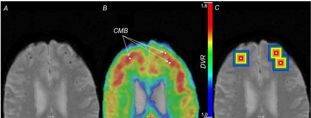
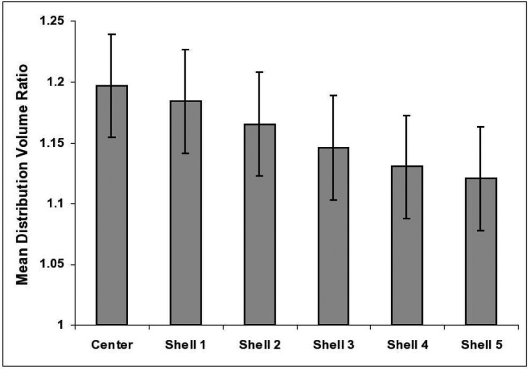
Advanced cerebrovascular β-amyloid deposition (cerebral amyloid angiopathy, CAA) is associated with cerebral microbleeds, but the precise relationship between CAA burden and microbleeds is undefined. We used T2*-weighted magnetic resonance imaging (MRI) and noninvasive amyloid imaging with Pittsburgh Compound B (PiB) to analyze the spatial relationship between CAA and microbleeds. On coregistered positron emission tomography (PET) and MRI images, PiB retention was increased at microbleed sites compared to simulated control lesions (p = 0.002) and declined with increasing distance from the microbleed (p < 0.0001). These findings indicate that microbleeds occur preferentially in local regions of concentrated amyloid and support therapeutic strategies aimed at reducing vascular amyloid deposition.
Figures
References
-
- Vinters HV, Gilbert JJ. Cerebral amyloid angiopathy: incidence and complications in the aging brain. II. The distribution of amyloid vascular changes. Stroke. 1983;14:924–928. - PubMed
-
- Rosand J, Muzikansky A, Kumar A, et al. Spatial clustering of hemorrhages in probable cerebral amyloid angiopathy. Ann. Neurol. 2005;58:459–462. - PubMed
-
- Klunk WE, Engler H, Nordberg A, et al. Imaging brain amyloid in Alzheimer's disease with Pittsburgh Compound-B. Ann. Neurol. 2004;55:306–319. - PubMed
-
- Johnson KA, Gregas M, Becker JA, et al. Imaging of amyloid burden and distribution in cerebral amyloid angiopathy. Ann. Neurol. 2007;62:229–234. - PubMed
Publication types
MeSH terms
Substances
Grants and funding
LinkOut - more resources
Full Text Sources
