

doi: 10.1002/ana.22138.
Direct interaction between causative genes of DYT1 and DYT6 primary dystonia
Affiliations
- PMID: 20865765
- PMCID: PMC3038652
- DOI: 10.1002/ana.22138
Item in Clipboard
Direct interaction between causative genes of DYT1 and DYT6 primary dystonia
Ann Neurol.
2010 Oct.
Abstract
Primary dystonia is a movement disorder characterized by sustained muscle contractions and in which dystonia is the only or predominant clinical feature. TOR1A(DYT1) and the transcription factor THAP1(DYT6) are the only genes identified thus far for primary dystonia. Using electromobility shift assays and chromatin immunoprecipitation (ChIP) quantitative polymerase chain reaction (qPCR), we demonstrate a physical interaction between THAP1 and the TOR1A promoter that is abolished by pathophysiologic mutations. Our findings provide the first evidence that causative genes for primary dystonia intersect in a common pathway and raise the possibility of developing novel therapies targeting this pathway.
Figures
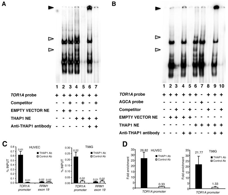
THAP1 binds the TOR1A promoter in vitro and in vivo. (A,B) Nuclear extracts (NE) from 293T cells transfected with wildtype human THAP1 were subjected to electrophoretic mobility shift assay (EMSA) using the TOR1A promoter region −159/−80 as a probe in A, or the same probe mutated in the THAP1 core consensus site (AGCA probe) in B. Unfilled triangles indicate specific nuclear complexes. Filled triangles indicate the supershifted bands. (C,D) ChIP-qPCR assays were used to analyze association of THAP1 with the TOR1A promoter in vivo. Cross-linked chromatin from HUVEC primary cells or T98G cells was subjected to immunoprecipitation with antibodies against THAP1 (THAP1 Ab1) or negative control antibodies (Control Ab). The human RRM1-exon 19 genomic region was used as a control genomic region. (C) Immunoprecipitated DNA was quantified by qPCR using the percent of input method. (D) Fold enrichment of THAP1 on the TOR1A promoter was calculated by dividing the amount of TOR1A promoter DNA precipitated by anti-THAP1 antibodies to the amount of DNA precipitated from the RRM1-exon 19 genomic region. ChIP results are shown as means with SD from three separate datapoints.
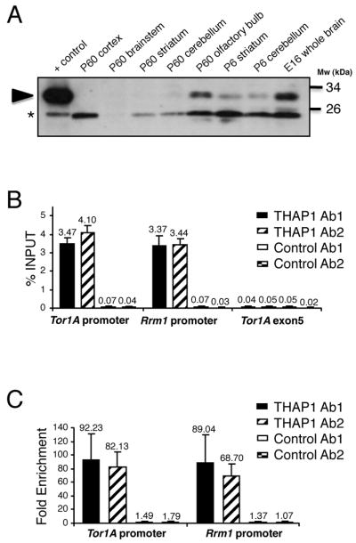
Endogenous THAP1 is bound to the Tor1A promoter in mouse brain. (A) Immunoblot analysis of THAP1 expression using anti-THAP Ab2 in nuclear fractions (80 μg/lane) of wildtype mouse brains structures obtained at indicated ages. Nuclear extracts (10 μg) of cells transfected with THAP1 were used as positive control. Arrowhead indicates band of the predicted size for THAP1. *Nonspecific immunoreactive band. (B,C) Cross-linked chromatin from adult mouse brain was subjected to immunoprecipitation with two distinct THAP1 (THAP1 Ab1 and THAP1 Ab2) and negative control antibodies (Control Ab1 and Control Ab2). The mouse Rrm1 promoter and Tor1A-exon 5 genomic region were used as positive and negative control regions, respectively. Immunoprecipitated DNA was quantified by qPCR (B) and fold enrichment (C) was calculated by dividing the amount of Tor1A or Rrm1 promoter DNA precipitated by anti-THAP1 antibodies to the amount of DNA precipitated from the Tor1A-exon 5 genomic region. Results are shown as means with SD from three separate datapoints.
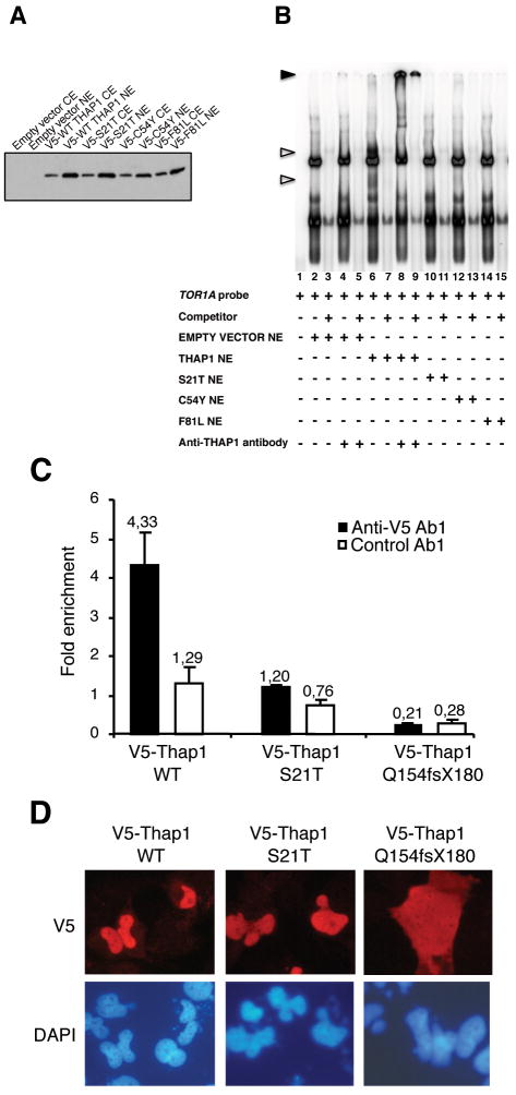
Causative mutations of DYT6 dystonia disrupt the THAP1/TOR1A interaction. (A) Immunoblot with monoclonal anti-V5 antibody showing similar levels of expression of the wildtype and mutant THAP1 proteins in cytoplasmic and nuclear extracts of 293T cells transfected with the indicated expression vectors. (B) Electrophoretic mobility shift assay (EMSA) showing that DYT6 patient point mutations disrupt physical interaction between THAP1 and TOR1A promoter region. Nuclear extracts from 293T cells transfected with the indicated expression vectors were used. Unfilled triangles indicate specific nuclear complexes. Filled triangles indicate the supershifted bands. (C,D) Cross-linked chromatin from T98G cells transfected with the indicated expression vectors was subjected to immunoprecipitation with anti-V5 or control antibodies. Immunoprecipitated DNA was quantified as described above (see Fig 1 legend) and fold enrichment of wildtype THAP1 or mutant proteins over the TOR1A promoter (C) was calculated by dividing the amount of TOR1A promoter DNA precipitated by anti-V5 antibodies to the amount of DNA precipitated from the RRM1-exon 19 genomic region. The results are shown as means ± SD from three separate datapoints. (D) Transfected T98G cells were analyzed by indirect immunofluorescence microscopy with anti-V5 antibodies (red). DNA was counterstained with DAPI (blue)
Comment in
-
A molecular link between dystonia 1 and dystonia 6?Ann Neurol. 2010 Oct;68(4):418-20. doi: 10.1002/ana.22183. Ann Neurol. 2010. PMID: 20976763 No abstract available.
References
-
- Fuchs T, Gavarini S, Saunders-Pullman R, et al. Mutations in the THAP1 gene are responsible for DYT6 primary torsion dystonia. Nat Genet. 2009;41:286–288. - PubMed
-
- Djarmati A, Schneider SA, Lohmann K, et al. Mutations in THAP1 (DYT6) and generalised dystonia with prominent spasmodic dysphonia: a genetic screening study. Lancet Neurol. 2009;8:447–452. - PubMed
Publication types
MeSH terms
Substances
Grants and funding
LinkOut - more resources
Full Text Sources
Other Literature Sources
