. 2010 Nov 2;49(43):9306-17.
doi: 10.1021/bi101336u.

Structure and mechanism of ORF36, an amino sugar oxidizing enzyme in everninomicin biosynthesis

Affiliations

Structure and mechanism of ORF36, an amino sugar oxidizing enzyme in everninomicin biosynthesis

Jessica L Vey et al. Biochemistry. .

Abstract

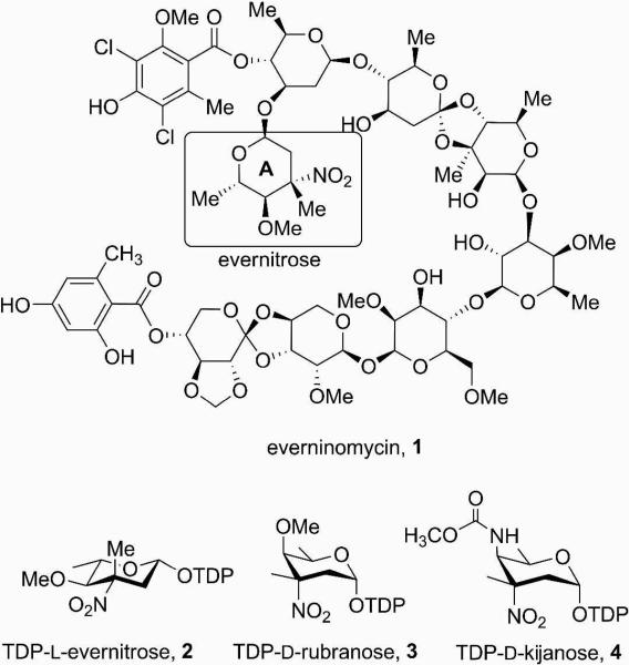
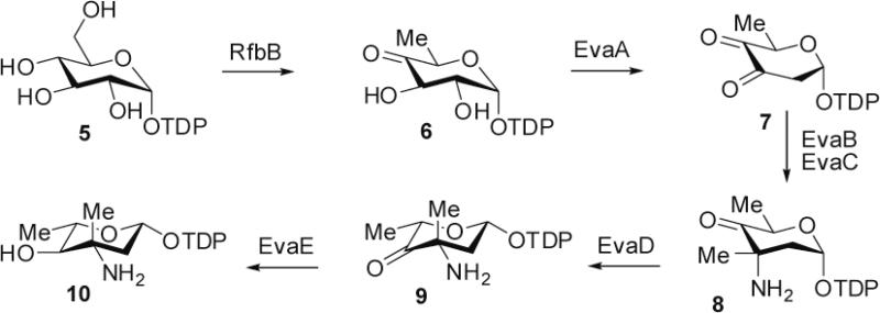
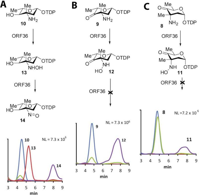
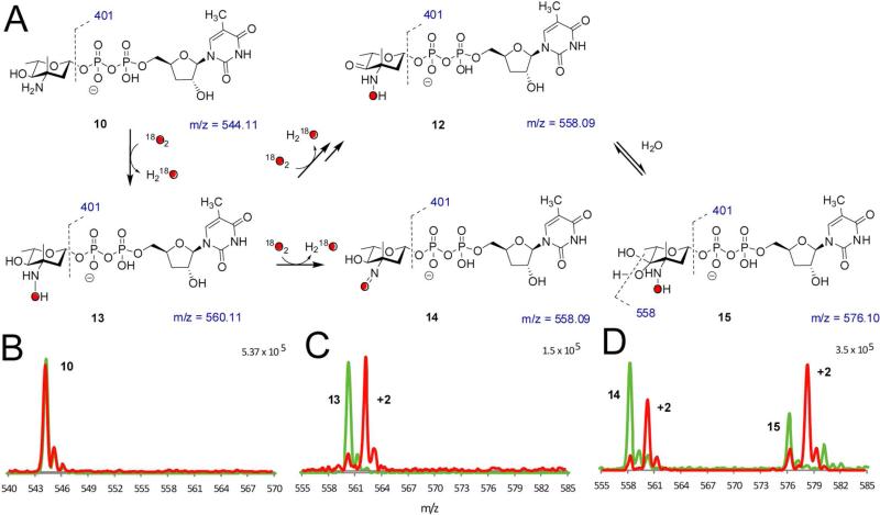
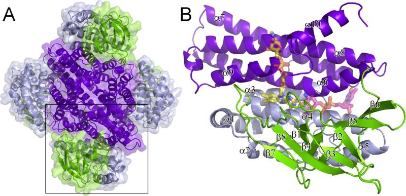
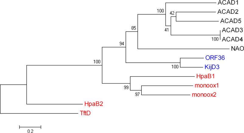
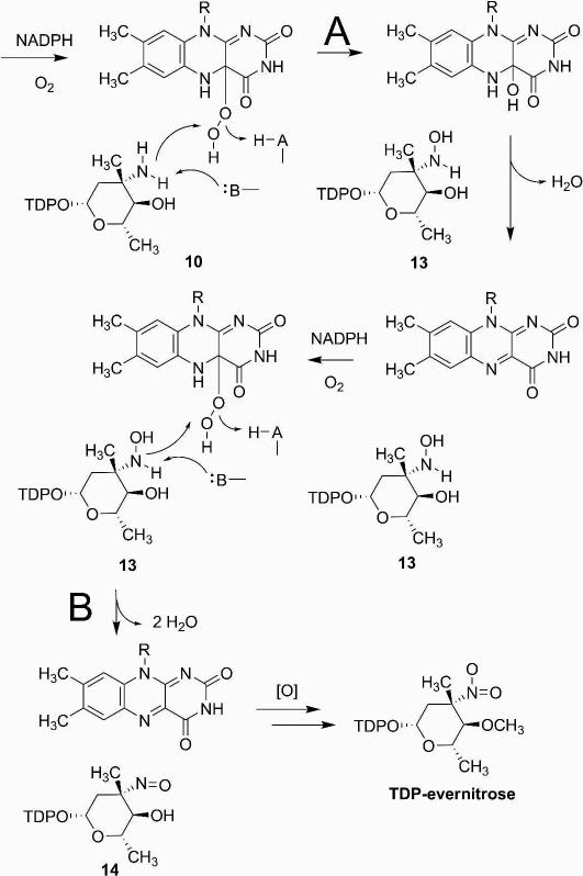
Everninomicin is a highly modified octasaccharide that belongs to the orthosomycin family of antibiotics and possesses potent Gram-positive antibiotic activity, including broad-spectrum efficacy against multidrug resistant enterococci and Staphylococcus aureus. Among its distinctive structural features is a nitro sugar, l-evernitrose, analogues of which decorate a variety of natural products. Recently, we identified a nitrososynthase enzyme encoded by orf36 from Micromonospora carbonacea var. africana that mediates the flavin-dependent double oxidation of synthetically generated thymidine diphosphate (TDP)-l-epi-vancosamine to the corresponding nitroso sugar. Herein, we utilize a five-enzyme in vitro pathway both to verify that ORF36 catalyzes oxidation of biogenic TDP-l-epi-vancosamine and to determine whether ORF36 exhibits catalytic competence for any of its biosynthetic progenitors, which are candidate substrates for nitrososynthases in vivo. Progenitors solely undergo single-oxidation reactions and terminate in the hydroxylamine oxidation state. Performing the in vitro reactions in the presence of (18)O(2) establishes that molecular oxygen, rather than oxygen from water, is incorporated into ORF36-generated intermediates and products and identifies an off-pathway product that correlates with the oxidation product of a progenitor substrate. The 3.15 Å resolution X-ray crystal structure of ORF36 reveals a tetrameric enzyme that shares a fold with acyl-CoA dehydrogenases and class D flavin-containing monooxygenases, including the nitrososynthase KijD3. However, ORF36 and KijD3 have unusually open active sites in comparison to these related enzymes. Taken together, these studies map substrate determinants and allow the proposal of a minimal monooxygenase mechanism for amino sugar oxidation by ORF36.


Figures

Figure 1
Figure 1
Everninomicin, its precursor TDP-l-evernitrose and related, proposed TDP-nitrosugar precursors to rubradirin and kijanimicin.
Figure 2
Figure 2
Pathway utilized for biochemical synthesis of TDP-l-epi-vancosamine 10 and keto amine precursors 8 and 9.
Figure 3
Figure 3
ORF36 catalysis with potential substrates. HPLC/ESI-MS traces of ORF36 reactions with three potential substrates. (A) TDP-l-epi-vancosamine 10 (m/z = 544), 0 min (blue), 100 min (green); hydroxylamine intermediate 13 (m/z = 560), 15 min (red); nitroso product 14 (m/z = 558), 100 min (purple). (B) keto amine 9 (m/z = 542), t = 0 min (blue), 100 min (green); hydroxylamine intermediate 12 (m/z = 558), 100 min (purple). (C) keto amine 8 (m/z = 542), 0 min (blue), 100 min (green); hydroxylamine intermediate 11 (m/z = 558), 100 min (purple).
Figure 4
Figure 4
18O2 incorporation studies. (A) Summary of proposed species observed in 18O2 incorporation experiments. (B – D) 18O2 incorporation with ORF36 and TDP-l-epi-vancosamine 10 incubated with 16O2 (green) and 18O2 (red). Masses shown under structures correspond to unlabeled species. HPLC/MS and MS/MS data were collected at a 15 minutes reaction time, when the hydroxylamine intermediate was most abundant. (B) TDP-l-epi-vancosamine 10 (tR = 4.5 min). (C) hydroxylamine 13 (m/z = 560, tR = 5.3 min). (D) Peaks at tR = 7 – 9 min are proposed to correspond to the nitroso compound 14 (tR = 8.8 min) and an additional oxidation product with m/z = 576 (tR = 7.8 min). This new mass has a distinct retention time from the nitrososugar 14 and, based on MS/MS analysis, the additional mass in the m/z = 576 ion is constrained to the pyranose ring. One possible structure is the 4-keto-3-hydroxylamino sugar 12, which is detected as a hydrate 15 under ESI conditions. MS/MS analysis supports this hypothesis in that the labeled ion m/z = 578 readily fragments to m/z = 560.09 and this fragmentation pattern is identical to the reaction product of 9 shifted by 2 mass units.
Figure 5
Figure 5
Structure of ORF36. (A) The ORF36 tetramer is shown as a cartoon representation with a transparent surface, colored by domain. The N-terminal domains (residues 1-129) are colored grey, the central β-sheet domains (residues 130-235) green and the C-terminal domains (residues 235-398) purple. (B) The ORF36 monomer shown in cartoon representation with modeled FAD and TDP-l-evernosamine shown in transparent sticks. Colors are as follows: FAD carbons, yellow; TDP-l-evernosamine carbons, magenta; oxygen, red; nitrogen, blue; phosphate, orange. The view in panel (B) is rotated 45° about a vertical axis from the bottom monomer shown boxed in A.
Figure 6
Figure 6
Evolutionary relationships of 13 taxa including nitrososynthases, acyl-CoA dehydrogenases, and flavin-containing monooxygenases. The evolutionary history was inferred using the Neighbor-Joining method (42). The bootstrap consensus tree inferred from 1000 replicates is taken to represent the evolutionary history of the taxa analyzed (43). Branches corresponding to partitions reproduced in less than 50% bootstrap replicates are collapsed. The percentage of replicate trees in which the associated taxa clustered together in the bootstrap test (1000 replicates) are shown next to the branches (43). The tree is drawn to scale, with branch lengths in the same units as those of the evolutionary distances used to infer the phylogenetic tree. The evolutionary distances were computed using the Poisson correction method (44) and are in the units of the number of amino acid substitutions per site. All positions containing gaps and missing data were eliminated from the dataset (Complete deletion option). There were a total of 333 positions in the final dataset. Phylogenetic analyses were conducted in MEGA4 (45). The nitrososynthases ORF36 and KijD3 are highlighted in blue text, the Class D flavin-containing monooxygenases in red, and the acyl-CoA dehydrogenases and nitroalkane oxidase are in black text. Proteins included here (which are also included in Supporting Information, Table S1) are named as follows: ACAD1, short branched-chain acyl-CoA dehydrogenase (H. sapiens), 2JIF (35); ACAD2, butyryl-CoA dehydrogenase (M. elsedenii), PDB entry 1BUC (58); ACAD3 medium-chain acyl-CoA dehydrogenase (S. scrofa), PDB entry 3MDE (59); ACAD4, medium-chain acyl-CoA dehydrogenase (S. scrofa), PDB entry 1UDY (60); ACAD5, short-chain acyl-CoA dehydrogenase (R. norvegicus), PDB entry 1JQI (61); HpaB1, 4-hydroxyphenylacetate monooxygenase (A. baumannii), PDB entry 2JBT (24); monoox1, 3-hydroxy-9,10-seco-nandrost-1,3,5(10)-triene-9,17-dione hydroxylase (Rhodococcus sp Rha1), PDB entry 2RFQ (62); monoox2, putative hydroxylase (Rhodococcus sp Rha1), PDB entry 2OR0 (63); NAO, nitroalkane oxidase (F. oxysporum), PDB entry 2C0U (64); HpaB2, 4-hydroxyphenylacetate monooxygenase (T. thermophilus), PDB entry 2YYI (25); TftD, chlorophenol-4-monooxygenase (B. cepacia), PDB entry 3HWC (26).
Figure 7
Figure 7
Active site loops. The loops that form the active site are as follows: the loops between α3 and α4 (loop L1; residues 106 – 109), β1 and β2 (loop L2; residues 134-142), β3 and β4 (loop L3, residues 158-164), β4 and β5 (loop L4; residues 178-180), β6 and β7 (loop L5; residues 201-213), β9 and α6 (loop L6; residues 248-253), α6’ and α7’ (where the ’ indicates this is from an adjacent monomer; loop L7; residues 272-280), α7 and α8 (loop L8; residues 310-317), α8’ and α9’ (loop L9, residues 351-353), α9 and α10, (loop L10; residues 375-379) and at the C-terminus (loop L11; residues 390-412). (A) A ribbon diagram of ORF36 shown with modeled flavin (yellow carbons) and TDP-l-evernosamine (magenta carbons) as sticks. Active site loops L3, L5, L7, and L9, are predicted to interact with flavin and are colored orange, active site loops L1, L4, L6, L8, and L11 are predicted to interact with substrate and are colored purple, and active site loops L2 and L10 are predicted to interact with both substrate and cofactor and are colored teal. (B) A ribbon diagram of human short branched-chain acyl Co-A dehydrogenase (PDB entry 2JIF) (35), highlighting loops L1 to L11 colored are colored as in (A). (C) A ribbon diagram of A. baumannii 4-hydroxyphenylacetate monooxygenase (PDB entry 2JBT) (24) highlighting loops L1 to L11 colored as in (A). (D) Stereoview of the ORF36 loop L10 containing a tandem cis-peptide. Loop L10 is shown in sticks with teal carbons. The Q376-P377 and P377-Y378 bonds both adopt a cis conformation. Modeled flavin and substrate are displayed as in (A).
Figure 8
Figure 8
Surface representation of the active site clefts of nitrososynthases, acyl-CoA dehydrogenases, and flavin-containing monooxygenases are shown colored by B-factor. Active site loops are labeled L1-L11. Surfaces of each enzyme are shown colored using a rainbow color ramp in which red corresponds to the highest B-factor and blue corresponds to the low B-factor. Areas of the surface that form the base of the active site cleft are shown in grey. Substrates and co-factors are shown as sticks with atoms colored as follows: FAD: carbons, yellow, oxygen, red; nitrogen, blue; phosphate, orange; TDP-l-evernosamine, dTDP, CoenzymeA persulfide: carbons, magenta; oxygen, red; nitrogen, blue; phosphate, orange. All panels are shown in an orientation rotated by 45° about a horizontal axis with respect to Figure 3A. (A) ORF36. Modeled FAD and TDP-l-evernosamine are displayed as sticks. In this orientation, loop L9 is concealed. The B factor color ramp gradient minimum and maximum values are 70 and 180 Å2, respectively, and for this structure, the Wilson B is 111.2 Å2 with an average protein B factor of 115.3 Å2. (B) KijD3 (PDB entry 3M9V)(11) with the dTDP of dTDP-phenol displayed as sticks. As in (A), loop L9 is concealed. The B factor color ramp minimum and maximum values for this panel are 10 and 80 Å2, and the average protein B value is 27.6 Å2. The Wilson B factor was not reported. (C) Human short/branched-chain acyl-CoA dehydrogenase (PDB entry 2JIF)(35). The FAD molecule is concealed by the protein surface in this orientation, and coenzymeA persulfide is shown in stick representation. Loops L9 and 10 are concealed in this orientation. The B factor color ramp gradient has minimum and maximum values of 14 and 60 Å2, the Wilson B is 25.84 Å2 and the average protein B factor is 22.76 Å2. (D) A. baumannii 4-hydroxyphenylacetate monooxygenase (PDB entry 2JBR)(24). FMN, 4-hydroxyphenylacetate, and loops L3, L9 and L10 are concealed in this orientation. The B factor color ramp gradient minimum and maximum values are 40 and 90 Å2, and the average protein B value is 56.77 Å2. The Wilson B factor was not reported.
Figure 9
Figure 9
Proposed monooxygenase mechanism of nitrososynthase. Pathway A denotes the first oxidation of substrate by ORF36, and pathway B shows the second oxidation step of the reaction cycle from substrate TDP-l-epi-vancosamine to the final nitrososugar product.

Similar articles

Cited by

References

    1. Ganguly AK. Ziracin, a novel oligosaccharide antibiotic. J. Antibiot. 2000;53:1038–1044. - PubMed
    1. Jones RN, Hare RS, Sabatelli FJ. In vitro Gram-positive antimicrobial activity of evernimicin (SCH 27899), a novel oligosaccharide, compared with other antimicrobials: a multicentre international trial. J. Antimicrob. Chemother. 2001;47:15–25. - PubMed
    1. Chu M, Mierzwa R, Jenkins J, Chan TM, Das P, Pramanik B, Patel M, Gullo V. Isolation and characterization of novel oligosaccharides related to Ziracin. J. Nat. Prod. 2002;65:1588–1593. - PubMed
    1. McNicholas PM, Najarian DJ, Mann PA, Hesk D, Hare RS, Shaw KJ, Black TA. Evernimicin binds exclusively to the 50S ribosomal subunit and inhibits translation in cell-free systems derived from both gram-positive and gram-negative bacteria. Antimicrob. Agents. Chemother. 2000;44:1121–1126. - PMC - PubMed
    1. Waitz JA, Horan AC. Micromonospora carbonacea var. africana. Schering Corporation; U.S.: 1988.

Publication types

MeSH terms

Associated data

LinkOut - more resources