Review
. 2010 Nov;391(11):1341-63.
doi: 10.1515/BC.2010.129.

Lipoprotein receptors--an evolutionarily ancient multifunctional receptor family

Affiliations
Review

Lipoprotein receptors--an evolutionarily ancient multifunctional receptor family

Marco Dieckmann et al. Biol Chem. 2010 Nov.

Abstract

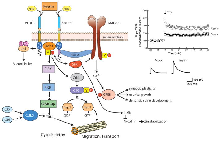
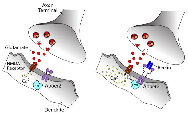
The evolutionarily ancient low-density lipoprotein (LDL) receptor gene family represents a class of widely expressed cell surface receptors. Since the dawn of the first primitive multicellular organisms, several structurally and functionally distinct families of lipoprotein receptors have evolved. In accordance with the now obsolete 'one-gene-one-function' hypothesis, these cell surface receptors were originally perceived as mere transporters of lipoproteins, lipids, and nutrients or as scavenger receptors, which remove other kinds of macromolecules, such as proteases and protease inhibitors from the extracellular environment and the cell surface. This picture has since undergone a fundamental change. Experimental evidence has replaced the perception that these receptors serve merely as cargo transporters. Instead it is now clear that the transport of macromolecules is inseparably intertwined with the molecular machinery by which cells communicate with each other. Lipoprotein receptors are essentially sensors of the extracellular environment that participate in a wide range of physiological processes by physically interacting and coevolving with primary signal transducers as co-regulators. Furthermore, lipoprotein receptors modulate cellular trafficking and localization of the amyloid precursor protein (APP) and the β-amyloid peptide (Aβ), suggesting a role in the pathogenesis of Alzheimer's disease. Moreover, compelling evidence shows that LDL receptor family members are involved in tumor development and progression.


Figures

Figure 1
Figure 1. The LDL receptor gene family
Panel A illustrates the core LDL receptor gene family as it exists in mammalian species. Panel B displays equivalent receptors that are structurally and functionally distinct family members in non-mammalian species. Panel C represents a subgroup of functionally important, but more distantly related family members that share some, but not all, of the structural requirements of the ‘core members’. In addition, they may also contain domains e.g. vacuolar protein sorting (VPS) domain, which are not present in the core family. These family members are characterized by one or more ligand binding domains, epidermal growth factor (EGF) – homology domains consisting of EGF repeats and YWTD propeller (β-propeller) domains involved in pH dependent release of ligands in the endosomes, a single transmembrane domain and a cytoplasmic tail containing at least one NPxY motifs. The latter represents both the endocytosis signal as well as a binding site for adaptor proteins linking the receptor to intracellular signaling pathways. Furthermore, LDLR, VLDLR, and Apoer2 carry an O-linked sugar domain.
Figure 2
Figure 2. Modulation of PDGF, TGFβ, and Wnt signaling activity by the LDL receptor family
Panel A, Regulation of PDGF signaling and suppression of atherosclerosis by LRP1. In the absence of LRP1, PDGF binds PDGF receptor β, induces its tyrosine phosphorylation, and activates PDGF-dependent migratory and proliferative signals. In the presence of LRP1, PDGF binds to both LRP1 and PDGF receptor β. Binding of ApoE-containing lipoproteins to LRP1 block LRP1 tail phosphorylation. In its unphosphorylated state, LRP1 interacts with adaptor proteins that are involved in the regulation of endocytosis (e.g. Dab2), reduces extracellular PDGF by endocytosis and degradation and prevents PDGF-dependent vascular smooth muscle cell migration and proliferation. In the absence of ApoE, LRP1 undergoes tyrosine phosphorylation in response to PDGF, which leads to recruitment of the Shc adaptor protein and activates proliferative and migratory signaling. Panel B, In the absence of LRP1 enhanced binding of cCbl to PDGFRβ results in increased monoubiquitination of PDGFRβ. This increases the affinity of the receptor for the ubiquitin interacting motifs in adaptor molecules such as Epsin/Eps15, and direct the receptor to coated pit endocytosis. LRP1 can interact with cCbl independently, which may competitively or sterically interfere with the association of cCbl with PDGFRβ, thereby decreasing the rate at which the receptor is ubiquitinated and removed from the cell surface by endocytosis. Panel C, Suppression of TGFβ and PDGF signaling and protection of vascular wall integrity by LRP1. Lack of LRP1 leads to enhanced complex formation of TGFβR1 and TGFβR2 and activation of TGFβ signaling resulting in phosphorylated SMAD2/3 (pSMAD2/3) accumulation, and increased PDGF receptor β expression and activation in smooth muscle cells. Moreover, LRP1 binds TGFβ directly or indirectly through α2M, internalizes insulin-like growth factor binding protein (IGFBP3) and the TGFβ activator thrombospondin (TSP), and sequesters TGFβ receptor. Panel D, Suppression of canonical Wnt signaling by LRP1 and LRP4. The ternary complex of Frizzled-1, co-receptor LRP5/6, and Wnt ligand at the cell surface leads to inhibition of GSK-3β and subsequent accumulation of β-catenin which translocates to the nucleus and mediates gene transcription. LRP1 and LRP4 suppress canonical Wnt signaling, probably by competing for LRP5/6 in the Wnt/Fz signaling complex at the membrane. In addition, LRP4 decreases Wnt signaling through its ligand binding domain by binding, sequestering or internalizing Wnt and BMP ligands and modulators. NPxY motif one formula image and two formula image.
Figure 3
Figure 3. Regulatory influence of LDLR family members on APP processing and Aβ production
APP bound to the cell surface is primarily processed by the α-secretase favoring the non-amyloidogenic pathway. Endocytosis of APP to the endosomal/lysosomal compartments results in increased β-secretase mediated cleavage and enhanced Aβ-production. The fast internalization rate of LRP1 facilitates APP endocytosis and its processing to Aβ. By contrast, LRP1B and Apoer2 endocytose slowly, which results in a longer residence time of APP at the cell surface promoting non-amyloidogenic processing. Apoer2 is associated with caveolae shifting APP from membrane non-raft to raft domains and thereby facilitating processing by β-secretase. SorLa prevents β-secretase access to APP and subsequent Aβ production by intracellularly shuttling APP to the Golgi compartment.
Figure 4
Figure 4. LRP1 mediates transcytosis of Aβ through the BBB
Cell surface LRP1 at the abluminal membrane binds Aβ and initiates its transcytosis across the BBB followed by its excretion into the blood stream. In addition, Aβ binding to LRP1 is influenced by Aβ transport proteins (ApoE, ApoJ, α2M, and lactoferrin). In the circulation, the soluble form of LRP1 binds >70% of Aβ and promotes its degradation.
Figure 5
Figure 5. Reelin signaling through VLDLR and Apoer2 in neurons (modified from (Herz et al. 2006))
Left Panel, The oligomeric signaling protein Reelin binds to VLDLR and Apoer2 with high affinity at the cell surface and induces receptor clustering and transactivation of Src family tyrosine kinases (SFKs), resulting in phosphorylation and thereby activation of disabled-1 (Dab1), an adaptor protein that interacts with NPxY motifs in both receptor tails. Phosphorylated Dab1 activates phosphatidylinositol 3-kinase (PI3K) and subsequently protein kinase B (PKB, Akt), which in turn inhibits glycogen synthase kinase-3β (GSK3β) and reduces phosphorylation of the microtubule stabilizing protein, tau. Tyrosine-phosphorylated Dab1 also recruit Crk and Crk-like (CrkL), which regulate actin cytoskeleton rearrangement through the phosphorylation of the guanine nucleotide exchange factor, C3G, and Rap1-GTP. Actin depolymerisation is also controlled by LIM kinase (LIMK), which inhibits N-cofilin by serine phosphorylation. Lis1 binds tyrosine-phosphorylated Dab1 and participates in the formation of a Pafah1b complex, which regulates microtubule functions. Cyclin-dependent kinase 5 (Cdk5) and its activators p35 and p39 are assumed to function in parallel with Reelin. In the synapse, Apoer2 associates with the post-synaptic scaffolding protein PSD95 through a 59 amino acid sequence encoded by the alternatively spliced exon 19, thus functionally coupling the Reelin signaling complex to the NMDA receptor (NMDAR). Reelin-activated SFKs phosphorylate the NMDAR on tyrosines in the NR2 subunits, thereby potentiating NMDAR-mediated Ca2+ influx. Elevated intracellular Ca2+ levels activate the transcription and survival factor cAMP-response element binding protein (CREB), which initiates the expression of genes that are important for synaptic plasticity, neurite growth and dendritic spine development. Right panel, Reelin potentiates long-term potentiation (LTP) (reproduced from (Weeber et al. 2002)) and enhances NMDAR-mediated whole cell current in wild type CA1 pyramidal neurons.
Figure 6
Figure 6. Reelin signaling through Apoer2 potentiates NMDAR-mediated Ca2+ influx at excitatory synapses
Ligation of Apoer2 by Reelin in the dendrite activates the SFK Fyn, which phosphorylates the NMDAR on NR2 subunits. Increased NMDAR phosphorylation stabilizes NMDAR at the plasma membrane (Snyder et al. 2005), and can also increase Ca2+ conductance. In this manner, the Reelin signaling pathway participates directly in the mechanisms that regulate synaptic plasticity, memory and learning.
Figure 7
Figure 7. LRP4 serves as a receptor for agrin and as a coreceptor for the tyrosine kinase MUSK in muscle
(Kim et al. 2008; Zhang et al. 2008). The membrane tyrosine kinase, MUSK, is required for the induction of acetylcholine receptor (AChR) clustering during the formation of neuromuscular junctions (endplates). MUSK does not bind the signaling protein agrin directly, but must form a complex with LRP4, a coreceptor for agrin. Agrin binding to LRP4 enhances complex formation of MUSK with LRP4 and induces transphosphorylation of MUSK followed by acetylcholine receptor (AChR) clustering.

References

    1. Andersen OM, Reiche J, Schmidt V, Gotthardt M, Spoelgen R, et al. Neuronal sorting protein-related receptor sorLA/LR11 regulates processing of the amyloid precursor protein. Proc Natl Acad Sci U S A. 2005;102:13461–6. - PMC - PubMed
    1. Andersen OM, Yeung CH, Vorum H, Wellner M, Andreassen TK, et al. Essential role of the apolipoprotein E receptor-2 in sperm development. J Biol Chem. 2003;278:23989–95. - PubMed
    1. Arnaud L, Ballif BA, Cooper JA. Regulation of protein tyrosine kinase signaling by substrate degradation during brain development. Mol Cell Biol. 2003;23:9293–302. - PMC - PubMed
    1. Arnaud L, Ballif BA, Forster E, Cooper JA. Fyn tyrosine kinase is a critical regulator of disabled-1 during brain development. Curr Biol. 2003;13:9–17. - PubMed
    1. Ashrafi K, Chang FY, Watts JL, Fraser AG, Kamath RS, et al. Genome-wide RNAi analysis of Caenorhabditis elegans fat regulatory genes. Nature. 2003;421:268–72. - PubMed

Publication types

MeSH terms