Review
. 2011 Jun 15;14(12):2545-79.
doi: 10.1089/ars.2010.3445. Epub 2011 Mar 10.

Apoptosis-inducing factor: structure, function, and redox regulation

Affiliations
Review

Apoptosis-inducing factor: structure, function, and redox regulation

Irina F Sevrioukova. Antioxid Redox Signal. .

Abstract

Apoptosis-inducing factor (AIF) is a flavin adenine dinucleotide-containing, NADH-dependent oxidoreductase residing in the mitochondrial intermembrane space whose specific enzymatic activity remains unknown. Upon an apoptotic insult, AIF undergoes proteolysis and translocates to the nucleus, where it triggers chromatin condensation and large-scale DNA degradation in a caspase-independent manner. Besides playing a key role in execution of caspase-independent cell death, AIF has emerged as a protein critical for cell survival. Analysis of in vivo phenotypes associated with AIF deficiency and defects, and identification of its mitochondrial, cytoplasmic, and nuclear partners revealed the complexity and multilevel regulation of AIF-mediated signal transduction and suggested an important role of AIF in the maintenance of mitochondrial morphology and energy metabolism. The redox activity of AIF is essential for optimal oxidative phosphorylation. Additionally, the protein is proposed to regulate the respiratory chain indirectly, through assembly and/or stabilization of complexes I and III. This review discusses accumulated data with respect to the AIF structure and outlines evidence that supports the prevalent mechanistic view on the apoptogenic actions of the flavoprotein, as well as the emerging concept of AIF as a redox sensor capable of linking NAD(H)-dependent metabolic pathways to apoptosis.


Figures

FIG. 1.
FIG. 1.
Major forms and splice variants of AIF. (A) Schematic representation of the human AIF gene. Exons are numbered; alternative exons giving splice variants are in gray. Translation initiation (ATG) and stop codons (TGA/TAA) are indicated. (B, E–G) Naturally occurring transcripts corresponding to the AIF precursor, AIFsh, AIFsh2, and AIFsh3, respectively. (C) Mature form of AIF produced upon mitochondrial processing. Depending on the usage of exons 2a and 2b, the ubiquitously expressed AIF1 or brain-specific AIF2 isoforms can be synthesized. (D) Truncated apoptogenic form produced in the intermembrane space upon proteolytic processing. The FAD-binding, NAD(H)-binding, and C-terminal (Ct) domains are indicated. AIF, apoptosis-inducing factor; MLS, mitochondrial leading sequence; NLS, nuclear leading sequence.
FIG. 2.
FIG. 2.
Phylogenetic tree of AIF-like proteins. Analysis of the phylogenetic relationships between the full-length molecular sequences was performed using the Phylogeny.fr online server (www.phylogeny.fr), which utilizes MUSCLE for sequence alignment and PhyML for phylogeny (55). GenBank or UniProt identification numbers are indicated.
FIG. 3.
FIG. 3.
Amino acid sequence alignment of AIF and AIF-like proteins from Homo sapiens (H. s.), Mus musculus (M. m.), Danio rerio (D. r.), Drosophila melanogaster (D. m.), Dictyostelium discoideum (D. d.), Caenorhabditis elegans (C. e.), Arabidopsis thaliana (A. t.), Pseudomonas sp. strain KKS102 (Ps.), and Saccharomyces cerevisiae (S. c.). Sequence alignment was performed with ClustalW2 (128). GenBank or UniProt accession numbers are given in Figure 2. Functionally important structural elements are highlighted in gray and indicated. The membrane-binding fragment in C. elegans is boxed.
FIG. 4.
FIG. 4.
Structure of AIF. (A) The x-ray model of refolded oxidized murine AIFΔ1–120 [PDB code 1GV4 (150)]. The FAD-binding, NAD(H)-binding, and C-terminal domains are depicted in beige, cyan, and purple, respectively; FAD is in CPK representation. The unique features of AIF are an extended N-terminus (shown in black), the N-terminal 190–202 insertion folded as a β-hairpin (deep cyan), and the C-terminal 509–559 insertion (red; proline residues are displayed). (B) Superposition of the crystallographic dimer of naturally folded oxidized murine AIFΔ1–77 [shown in gray, PDB code 3GD3 (202)] and biological dimer of the reduced NAD-bound AIFΔ1–101 [brown, PDB code 3GD4 (202)]. FAD is in CPK representation; NAD is in green. (C) A hydrogen-bonding network in the active site of reduced AIF. The nicotinamide group (green) is parallel stacked between Phe309 and the isoalloxazine of FAD (yellow) and establishes H-bonding network with the surrounding residues. One of these, His453, undergoes a large positional shift upon AIF reduction. (D, E) Views at the front side and the top of the reduced AIF dimer, respectively. The redox-sensitive 190–202 β-hairpin and Trp195 (in CPK representation) are shown in blue. NLS1 and NLS2 are in pink and magenta, respectively. NLS2, through which AIF is predominantly transported to the nucleus, comprises the dimer interface and, hence, becomes inaccessible upon AIF reduction. The α-helical portion of the regulatory peptide unwinds, and the 536–543 amino acid stretch transforms into the sixth strand of the C-terminal β-sheet (shown in red). The remaining residues of the regulatory peptide are not seen in the x-ray structure due to disorder (To see this illustration in color the reader is referred to the web version of this article at www.liebertonline.com/ars).
FIG. 5.
FIG. 5.
Structural alterations in AIF caused by refolding. (A) Conformational differences in FAD and the 438–453 peptide, spanning from the crystallographic dimer interface to the active site. Due to conformational differences in the side chain of Arg448, forming a salt bridge with Asp414 in refolded AIF (gray) and an H-bond with the Val422 carbonyl oxygen in the naturally folded protein (black), the 438–540 fragment folds as a loop or β-turn, respectively. (B) By establishing an intersubunit salt bridge with Glu412, Arg448 may assist dimerization and stabilization of NADH-reduced AIF. Conformational alterations in the 438–453 peptide detected in refolded AIF (displayed in gray) could interfere with the dimerization process and be responsible for the perturbed redox properties of the protein.
FIG. 6.
FIG. 6.
Extension of the C-terminal antiparallel β-sheets in crystalline AIFΔ1–77. The N-termini of the neighboring molecules forming β-strands are shown in black and indicated by arrows.
FIG. 7.
FIG. 7.
Redox-induced reorganization of aromatic residues in the C-terminal domain of murine AIFΔ1–101. Displacement of Phe481 and Phe309 by the nicotinamide group initiates the rearrangement and leads to formation of an aromatic tunnel that may serve as an electron delocalization site and prolong the lifetime of the reduced species.
FIG. 8.
FIG. 8.
Amino acid composition of the regulatory peptide in human and murine AIF (upper and lower sequences, respectively). The proline-rich motif within the PEST sequence is boxed; two lysines critical for DNA-binding are shown in bold italic. The putative phosphorylation sites were identified using the online NetPhos server (www.cbs.dtu.dk/services/NetPhos/) and are marked by asterisks. Human AIF has one additional phosphorylation site at Thr547, which is substituted by Ala in the murine protein. An arrow indicates the peptide transforming into the sixth strand of the C-terminal β-sheet upon AIF reduction.
FIG. 9.
FIG. 9.
Factors modulating the cleavage and release of apoptogenic AIF from mitochondria. Proteolysis of the membrane tether in mature AIF can be mediated by local or cytoplasmic proteases entering the intermembrane space upon permeabilization of the outer membrane. Detached AIF can translocate to the cytoplasm with the involvement of the PTP complex or through pores formed by proapoptotic Bcl-2 family members Bax, Bak, and Bid. AIF associated with the outer leaflet of the OMM, likely a full-length precursor, can be released and transported to the nucleus in a PAR-dependent manner. ANT, adenine nucleotide translocase; PAR, poly(ADP-ribose); PTP, permeability transition pore; VDAC, voltage-dependent anion channel.
FIG. 10.
FIG. 10.
Cytoplasmic partners of apoptogenic AIF. After release into the cytoplasm, AIF can promote apoptosis by interacting with TULA, eIF3g, and phospholipid scramblase, the pro-death partners. Scythe facilitates cell death by regulating stability and lifetime of the cytoplasmic AIF precursor, wherein CypA assists cytonuclear translocation of apoptogenic AIF. Contrarily, Hsp70 retains AIF in the cytoplasm and, hence, can postpone or prevent initiation of the nuclear apoptosis. XIAP is another pro-life partner, which in co-operation with AIF reduces reactive oxygen species levels and promotes cell survival. CypA, cyclophilin A; eIF3g, eukaryotic translation initiation factor 3 subunit p44; Hsp70, 70 kDa heat shock protein; TULA, T-cell ubiquitin ligand; XIAP, X-linked inhibitor of apoptosis protein.
FIG. 11.
FIG. 11.
An Hsp70 binding site identified by systematic deletion analysis and in silico modeling (84, 196). The 150–228 and, more precisely, 184–221 fragment (shown in dark gray and cartoon representation) and Arg192 and Lys194 in human AIF were identified as important for Hsp70 binding. Since the predicted site includes a redox-sensitive 191–203 β-hairpin, interaction between AIF and Hsp70 may be redox controlled.
FIG. 12.
FIG. 12.
Complexes between the BIR2 domain of human XIAP (PDB code 1I30) and the oxidized monomer and reduced dimer of AIF (A and B, respectively) generated with the program GRAMM (220). AIF molecules are in light gray and cartoon representation; BIR2 is in black and ribbon representation. In both types of complexes, the top ranking solutions for BIR2 are clustered at two docking sites that have similar binding energy. This suggests that association of AIF with XIAP may be redox independent.
FIG. 13.
FIG. 13.
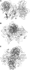
Complexes between CypA (PDB code 3KOM) and the oxidized monomer (A) and reduced dimer of AIF (B, C) generated with the program GRAMM (220). AIF molecules are depicted in light gray and carton representation. Computer modeling suggests that CypA (in ribbon representation) has a higher affinity for the dimeric form of AIF and preferably binds to the grove above the monomer–monomer interface. In the oxidized monomer, two favorable CypA docking positions are predicted, distinct from the site identified by Cande et al. [residues 367–399 (29); shown in black and indicated by an arrow]. Physiological relevance of the predicted CypA binding sites and whether the AIF-CypA interaction is redox dependent or simply requires clustering and oligomerization of AIF remain to be established.
FIG. 14.
FIG. 14.
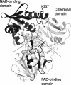
A potential binding site for phospholipid scramblase derived based on the sequence homology between human AIF and C. elegans WAH-1 (231). The 270–440 fragment and Lys337 in human AIF (highlighted in black) correspond to the WAH-1 380–550 peptide and Lys446, which are critical for interaction with SCRM-1, a worm homolog of human scramblase. The predicted scramblase-binding site includes residues comprising the NAD(H)- and FAD-binding domains.
FIG. 15.
FIG. 15.
Nuclear effects of apoptogenic AIF. AIF is transported from the cytoplasm to the nucleus by nuclear transport receptors recognizing NLS1 and NLS2. CypA assists and Hsp70 prevents cytonuclear translocation of AIF. In the nucleus, AIF directly binds to DNA and in cooperation with other proteins causes peripheral chromatin condensation and, possibly, large-scale chromatin fragmentation. Histone H2AX and CypA cooperatively promote AIF-mediated nuclear apoptosis. Co-operation between AIF and endonuclease G was suggested but has not been proven thus far. Other unidentified proteins (indicated in the figure as triangles with a question mark) may interact with AIF as well, forming a DNA degrading complex, “degradosome.” Upon chromatinolysis, AIF might dissociate from oligonucleosomal DNA and translocate back to the cytoplasm.
FIG. 16.
FIG. 16.
(A) The C-terminal sequence alignment of the AIF-like proteins and (B) the x-ray structure of human AIF. (A) A DNA-binding helix-turn-helix motif predicted in D. discoideum is boxed. Residues corresponding to the 512Lys-Arg-Arg514 cluster in D. discoideum are also indicated. (B) The 595–605 peptide corresponding to the DNA-binding motif in D. discoideum is displayed in black. Instead of the basic cluster predicted to comprise the turn between helices in D. discoideum, human AIF has 594Asp-Gly-Glu596, which makes the surface more acidic and less suitable for DNA binding.
FIG. 17.
FIG. 17.
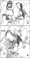
Functional importance of Arg201. (A) Superposition of the x-ray structures of oxidized human and murine AIF. By forming a salt bridge with Glu531, Arg201 assists folding of the β-hairpin and the helical part of the regulatory peptide (highlighted in black), which stabilizes FAD binding. (B) Superposition of the reduced forms of wild-type (light gray) and ΔR201 mutant (black) of human AIF, modeled based on the structure of the murine protein. In the wild-type protein, Arg201 forms an H-bond with the carbonyl oxygen of Phe204 and limits the flexibility of the β-hairpin. Deletion of Arg201 shortens the hairpin and disrupts the β-turn structure. In oxidized AIF this may lead to weaker FAD association, whereas in the reduced form, where binding of FAD is stabilized through charge–transfer and π-π stacking interactions with NAD, the Arg201 deletion and elimination of the H-bond with Phe204 may increase flexibility of the partially unstructured 191–203 peptide and, possibly, perturb protein–protein interactions. Through these changes, the ΔR201 mutation can affect both vital and apoptogenic functions of AIF.
FIG. 18.
FIG. 18.
Possible redox-sensing mechanism of AIF. In normal mitochondria, the reduced dimer is likely to be the predominant form of AIF and could assist cristae formation and OXPHOS functioning through a specific redox activity or/and protein–protein interactions. Fluctuations in the NAD(H) levels and aberrations in the inner membrane could lead to transient changes in monomer–dimer equilibrium and dissociation of the AIF-mediated protein complexes (preapoptotic state). Depletion of pyridine nucleotides and severe defects in the inner membrane coupled with the protease activation and OMM permeabilization would lead to AIF monomerization, proteolysis, and translocation into the cytoplasm. Owing to the distinctly folded β-hairpin and regulatory peptides (shown as bold lines) and differences in the accessibility of NLS2, the oxidized and reduced forms of AIF could recruit different cytoplasmic partners and, consequently, initiate different signaling pathways. The loss of redox active mitochondrial AIF, leading to OXPHOS failure and increased production of reactive oxygen species, may be another factor promoting cell death. If the reduced dimer does relocate to the nucleus, it will be less apoptogenic than the oxidized monomer due to the lower affinity for DNA. Existence of separate mitochondrial, cytoplasmic, and nuclear NAD(H) pools allows compartmental regulation of the AIF-mediated signal transduction and creates multilevel check points through which the cell can reverse, postpone, or promote its demise. OXPHOS, oxidative phosphorylation.

Similar articles

Cited by

References

    1. Ahel I. Ahel D. Matsusaka T. Clark AJ. Pines J. Boulton SJ. West SC. Poly(ADP-ribose)-binding zinc finger motifs in DNA repair/checkpoint proteins. Nature. 2008;451:81–85. - PubMed
    1. Akematsu T. Endoh H. Role of apoptosis-inducing factor (AIF) in programmed nuclear death during conjugation in Tetrahymena thermophila. BMC Cell Biol. 2010;11:13. - PMC - PubMed
    1. Alano CC. Garnier P. Ying W. Higashi Y. Kauppinen TM. Swanson RA. NAD+ depletion is necessary and sufficient for poly(ADP-ribose) polymerase-1-mediated neuronal death. J Neurosci. 2010;30:2967–2978. - PMC - PubMed
    1. Anderson P. Kedersha N. Stress granules: the Tao of RNA triage. Trends Biochem Sci. 2008;33:141–150. - PubMed
    1. Andrabi SA. Kim NS. Yu SW. Wang H. Koh DW. Sasaki M. Klaus JA. Otsuka T. Zhang Z. Koehler RC. Hurn PD. Poirier GG. Dawson VL. Dawson TM. Poly(ADP-ribose) (PAR) polymer is a death signal. Proc Natl Acad Sci U S A. 2006;103:18308–18313. - PMC - PubMed

MeSH terms

LinkOut - more resources