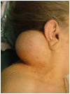
Hemangiopericytoma of the neck
- PMID: 20868476
- PMCID: PMC2954839
- DOI: 10.1186/1746-160X-6-23
Hemangiopericytoma of the neck
Abstract
Hemangiopericytoma (HPC) is an exceedingly rare tumor of uncertain malignant potential. Approximately 300 cases of HPC have been reported since Stout and Murray described HPCs as "vascular tumors arising from Zimmerman's pericytes" in 1942. After further characterization, the WHO reclassified HPC as a fibroblastic/myofibroblastic tumor. Long term follow up is mandatory because the histologic criteria for prediction of biologic behavior are imprecise. There are reports of recurrence and metastasis many years after radical resection. The head and neck incidence is less than 20%, mostly in adults. We report herein a case of HPC resected from the neck of a 74-year-old woman, who presented in our department with a painless right-sided neck mass. The mass was well circumscribed, mobile and soft during the palpation. The skin over the tumor was intact and normal. Clinical diagnosis at this time was lipoma. A neck computer tomography scan showed a large submucosal mass in the neck, which extended in the muscular sites. The tumor was completely removed by wide surgical resection. During surgery we found a highly vascularised tumor. The histopathologic examination revealed a cellular, highly vascularized tumor. The diagnosis was that of solitary fibrous tumor, cellular variant, with haemangiopericytoma-like features. The patient had normal postoperative course of healing and 24 months later she remains asymptomatic, without signs of recurrence or metastases.
Figures


References
-
- Wagner E, Wunderlich CA, Roser W. Das tuberkelähnliche Lymphadenom. (Hrsg) 1870. pp. S497–525.
Publication types
MeSH terms
LinkOut - more resources
Full Text Sources
Medical

