Review
. 2010 Oct;9(5):237-55.

Image-guided surgery using invisible near-infrared light: fundamentals of clinical translation

Affiliations
Review

Image-guided surgery using invisible near-infrared light: fundamentals of clinical translation

Sylvain Gioux et al. Mol Imaging. 2010 Oct.

Abstract

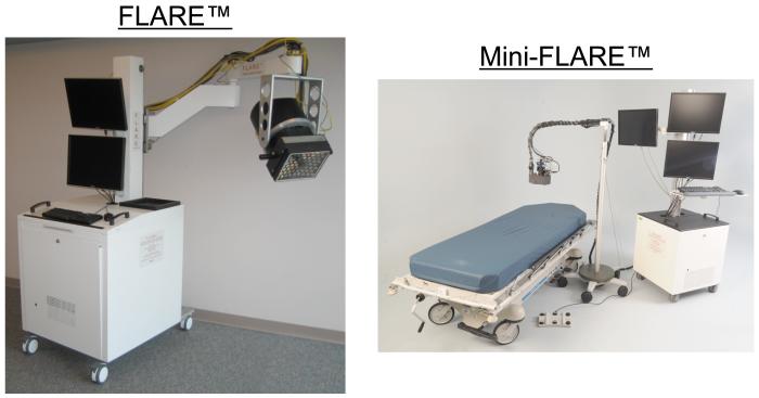
The field of biomedical optics has matured rapidly over the last decade and is poised to make a significant impact on patient care. In particular, wide-field (typically > 5 cm), planar, near-infrared (NIR) fluorescence imaging has the potential to revolutionize human surgery by providing real-time image guidance to surgeons for tissue that needs to be resected, such as tumors, and tissue that needs to be avoided, such as blood vessels and nerves. However, to become a clinical reality, optimized imaging systems and NIR fluorescent contrast agents will be needed. In this review, we introduce the principles of NIR fluorescence imaging, analyze existing NIR fluorescence imaging systems, and discuss the key parameters that guide contrast agent development. We also introduce the complexities surrounding clinical translation using our experience with the Fluorescence-Assisted Resection and Exploration (FLARE™) imaging system as an example. Finally, we introduce state-of-the-art optical imaging techniques that might someday improve image-guided surgery even further.


Figures

Figure 1
Figure 1. FLARE™ and Mini-FLARE™ Image-Guided Surgery Systems
The FLARE™ imaging system (left) is composed of a cart containing control electronics, cooling, and a computer, an articulating providing 6 degrees of freedom, 127 cm lateral reach, and 178 cm vertical reach, and an imaging head containing a custom high power LED light source, 2 NIR and 1 color cameras, and custom optics. The Mini-FLARE™ imaging system (right; Troyan et al., manuscript in review) represents a significant reduction in size and improvement in flexibility.
Figure 2
Figure 2. Contrast Agents for NIR Fluorescence-Guided Surgery
  1. General structure of the heptamethine indocyanine class of NIR fluorophores.

  2. Chemical structures and key optical properties (in serum) of the clinically available NIR fluorophores methylene blue (MB; left) and indocyanine green (ICG; right).

Figure 3
Figure 3. FLARE™ Imaging System Optical Channels in the Context of Clinically-Available NIR Fluorophores
The color video, 700 nm NIR fluorescence and 800 nm NIR fluorescence optical channels of the FLARE™ imaging system are shown, along with the absorbance (left axis) and fluorescence (right axis) of 5 μM methylene blue (MB) and 2.5 μM indocyanine green (ICG) in 100% serum, pH 7.4.
Figure 4
Figure 4. Concentration-Dependent Quenching of MB and ICG using Different Optical Geometries
NIR excitation and emission geometries were either 0° or 90° as indicated (top). Shown at bottom is the concentration-dependent quenching of methylene blue (MB) and indocyanine green (ICG) in 100% fetal bovine serum supplemented with 50 mM HEPES, pH 7.4 under conditions of 0° (open circles) or 90° (closed squares) excitation/emission geometries.
Figure 5
Figure 5. Clinical Imaging using the FLARE™ Imaging System
A. Sentinel lymph node (arrow) mapping in the axilla of a woman with breast cancer after peri-tumoral injection of ICG:HSA as described in . NIR fluorescence = 800 nm channel; 67 msec exposure time. The NIR fluorescence image was pseudo-colored in lime green and superimposed on the color video image to produce Color-NIR Merge. B. Perforator flap NIR fluorescence angiography after intravenous injection of ICG. Dominant perforator artery (dashed circle) identified during dynamic imaging (Lee et al., manuscript in review). NIR fluorescence = 800 nm channel; 67 msec exposure time. The NIR fluorescence image was pseudo-colored in lime green and superimposed on the color video image to produce Color-NIR Merge.

References

    1. Frangioni JV. New technologies for human cancer imaging. J Clin Oncol. 2008;26:4012–21. - PMC - PubMed
    1. Bani MR, Lux MP, Heusinger K, et al. Factors correlating with reexcision after breast-conserving therapy. Eur J Surg Oncol. 2008 - PubMed
    1. Schiller DE, Le LW, Cho BC, et al. Factors associated with negative margins of lumpectomy specimen: potential use in selecting patients for intraoperative radiotherapy. Ann Surg Oncol. 2008;15:833–42. - PubMed
    1. Besana-Ciani I, Greenall MJ. The importance of margins status after breast conservative surgery and radiotherapy in node positive patients: a follow-up of 10-15 years. Int Semin Surg Oncol. 2008;5:13. - PMC - PubMed
    1. Burke S, Shorten GD. When pain after surgery doesn’t go away. Biochem Soc Trans. 2009;37:318–22. - PubMed

Publication types