Review
. 2011 Jan;30(1):18-53.
doi: 10.1016/j.preteyeres.2010.09.001. Epub 2010 Sep 22.

Dietary hyperglycemia, glycemic index and metabolic retinal diseases

Affiliations
Review

Dietary hyperglycemia, glycemic index and metabolic retinal diseases

Chung-Jung Chiu et al. Prog Retin Eye Res. 2011 Jan.

Abstract

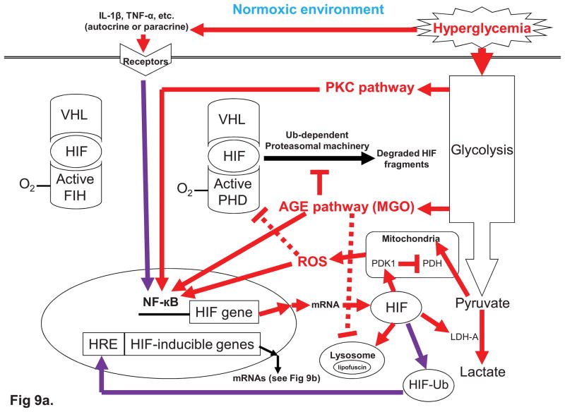
The glycemic index (GI) indicates how fast blood glucose is raised after consuming a carbohydrate-containing food. Human metabolic studies indicate that GI is related to patho-physiological responses after meals. Compared with a low-GI meal, a high-GI meal is characterized with hyperglycemia during the early postprandial stage (0-2h) and a compensatory hyperlipidemia associated with counter-regulatory hormone responses during late postprandial stage (4-6h). Over the past three decades, several human health disorders have been related to GI. The strongest relationship suggests that consuming low-GI foods prevents diabetic complications. Diabetic retinopathy (DR) is a complication of diabetes. In this aspect, GI appears to be useful as a practical guideline to help diabetic people choose foods. Abundant epidemiological evidence also indicates positive associations between GI and risk for type 2 diabetes, cardiovascular disease, and more recently, age-related macular degeneration (AMD) in people without diabetes. Although data from randomized controlled intervention trials are scanty, these observations are strongly supported by evolving molecular mechanisms which explain the pathogenesis of hyperglycemia. This wide range of evidence implies that dietary hyperglycemia is etiologically related to human aging and diseases, including DR and AMD. In this context, these diseases can be considered as metabolic retinal diseases. Molecular theories that explain hyperglycemic pathogenesis involve a mitochondria-associated pathway and four glycolysis-associated pathways, including advanced glycation end products formation, protein kinase C activation, polyol pathway, and hexosamine pathway. While the four glycolysis-associated pathways appear to be universal for both normoxic and hypoxic conditions, the mitochondria-associated mechanism appears to be most relevant to the hyperglycemic, normoxic pathogenesis. For diseases that affect tissues with highly active metabolism and that frequently face challenge from low oxygen tension, such as retina in which metabolism is determined by both glucose and oxygen homeostases, these theories appear to be insufficient. Several lines of evidence indicate that the retina is particularly vulnerable when hypoxia coincides with hyperglycemia. We propose a novel hyperglycemic, hypoxia-inducible factor (HIF) pathway, to complement the current theories regarding hyperglycemic pathogenesis. HIF is a transcription complex that responds to decrease oxygen in the cellular environment. In addition to playing a significant role in the regulation of glucose metabolism, under hyperglycemia HIF has been shown to increase the expression of HIF-inducible genes, such as vascular endothelial growth factor (VEGF) leading to angiogenesis. To this extent, we suggest that HIF can also be described as a hyperglycemia-inducible factor. In summary, while management of dietary GI appears to be an effective intervention for the prevention of metabolic diseases, specifically AMD and DR, more interventional data is needed to evaluate the efficacy of GI management. There is an urgent need to develop reliable biomarkers of exposure, surrogate endpoints, as well as susceptibility for GI. These insights would also be helpful in deciphering the detailed hyperglycemia-related biochemical mechanisms for the development of new therapeutic agents.


Figures

Fig. 1
Fig. 1
Adverse metabolic events relating high-GI diets to diabetes and cardiovascular disease.
Fig. 2
Fig. 2
Glycemic responses demonstrate the definition of GI.
Fig. 3
Fig. 3
Studies relating GI to AMD indicate that consuming a low-GI diet is associated with lower risk for both early and advanced AMD.
Fig. 4
Fig. 4. Cellular responses to euglycemia (normal glycemia) and hyperglycemia under normoxia (4a) and hypoxia (4b)
Fig. 4a. Glucose metabolism in euglycemia vs. hyperglycemia under normoxic conditions. Compared with euglycemia, hyperglycemia induces mitochondria-derived superoxide (O2) and four glycolysis-related pathways (see Figs 5–8), including polyol, hexosamine, AGE, and PKC pathways, and excess cytosolic HIF. The left panel demonstrates normal aerobic respiration in a euglycemic condition. After glycolysis, the glucose metabolite, pyruvate, is produced. Pyruvate enters the mitochondria to generate ATP and water (H2O). The right panel demonstrates that hyperglycemia drives glycolysis to generate the four adverse side pathways noted above.(also see Fig. 5–8) In the mean time, driven by the hyperglycemia, the ETC is obstructed in coenzyme Q by an abnormally high mitochondrial membrane potential and generates superoxide (O2), which may activate PARP, a DNA repair enzyme which needs GAPDH as a cofactor and is only found in the nucleus. This gives rise to the decrease of cytosol GAPDH and further exacerbates of the four glycolysis-associated pathways induced by hyperglycemia. The mechanism underlying the movement of GAPDH from the cytosol to the nucleus under high glucose conditions involves the E3 Ub ligase siah-1, which facilitates hyperglycemia-induced GAPDH nuclear translocation via formation of a complex with GAPDH. Furthermore, because hyperglycemic AGEs, PKC, and mitochondrial ROS may give rise to the over expression and decreased degradation of HIF, the excess HIF proteins may switch pyruvate metabolism from transformation through the TCA cycle and oxidative phosphorylation in the ETC to conversion to lactate in the cytoplasm (also see Fig. 9a for more details). Remarkablly, PKC can be also activated through hyperglycemic polyol pathway (Fig. 6) and hyperglycemic hexosamine pathway (Fig. 8). The cell may defend against superoxide using the mitochondrial isoform of superoxide dismutase (Mn-SOD). This enzyme degrades the oxygen free radical to hydrogen peroxide, which is then converted to H2O and O2 by other enzymes. Fig. 4b. Glucose metabolism in euglycemia vs. hyperglycemia under hypoxic conditions. In euglycemia, HIF pathway is turn on by hypoxia-activated HIF. Under hyperglycemic conditions, the HIF pathway is enhanced by hyperglycemia-induced AGE and PKC pathways. The left panel indicates the HIF is activated and induces two aspects of the cellular responses, including switching glucose metabolism and turning on HIF pathway. Cytosolic HIF switches glucose metabolism from aerobic respiration to fermentation, the end product of which is lactate. The HIF pathway activated by hypoxia-activated HIF may induce a range of deleterious effects. However, when hypoxia coincides with hyperglycemia (right panel), which results in the formation AGEs and activation of PKC during glycolysis, HIF pathway is further enhanced by hyperglycemia. Remarkablly, PKC can be also activated through hyperglycemic polyol pathway (Fig. 6) and hyperglycemic hexosamine pathway (Fig. 8). Furthermore, in adaptation of lower efficiency of ATP generation from fermentation, the activations of some HIF-inducible genes in HIF pathway may increase glucose uptake and up-regulate glycolysis pathway (also see Fig. 9b). Therefore, in hyperglycemic, hypoxic conditions HIF pathway may further deteriorate the four glycolysis-associated pathways.
Fig. 5
Fig. 5. Hyperglycemic AGE pathway
The hyperglycemia-induced intracellular AGE precursors, such as MGO, induce pathological consequences in four routes, 1) direct intracellular glycation of proteins, including proteins involved in the regulation of gene transcription, such as NF-κB, 2) inhibiting enzymes responsible for protein degradation, such as proteasomal (including ubiquitin) and lysosomal systems, 3) the intracellular AGEs precursors can diffusing out of the cell and modify nearby cells (even the same cell itself), extracellular matrix, such as Bruch’s membrane and choroidal capillary membranes, and 4) the intracellular AGEs precursors diffusing out of the cell to modify circulating proteins in the blood, which in turn activate RAGE on pro-inflammatory cells or CECs, thereby causing the production of inflammatory cytokines and/or growth factors.
Fig. 6
Fig. 6. Hyperglycemic polyol pathway
Under hyperglycemia, AR reduces glucose to sorbitol (a polyol or sugar alcohol), which is later oxidized to fructose. In this process, the AR consumes cofactor NADPH. Therefore, the hyperglycemic polyol pathway consumes NADPH and hence results in the depletion of GSH. This increases intracellular oxidative stress.
Fig. 7
Fig. 7. Hyperglycemic PKC pathway
The pathogenic consequences of hyperglycemic PKC through activating transcription factors for a wide range of proteins, including cytokines. Many transcription factors, such as NF-κB, are activated through hyperglycemia-induced PKC activation, resulting in oxidatives stress, increased vaso-permeability, angiogenesis, vascular occlusion, capillary occlusion, and abnormal blood flow, etc.
Fig. 8
Fig. 8. Hyperglycemic hexosamine pathway
The hyperglycemic hexosamine pathway starting from the glycolytic intermediate, F-6-P, which is converted by GFAT to glucosamine-6-P and eventually to UDPGlcNAc, an O-linked GlcNAc. Intracellular glycosylation by adding GlcNAc moieties to serine and threonine residues of proteins (e.g. transcription factors) is catalysed by OGT. Increased glycosylation of transcription factors, such as Sp1, AP2 and CREB, often at phosphorylation sites, increases the expression of cytokines and enzymes, including TGF- β 1, PAI-1, and glycosyltransferase. In addition, AGEs can exert cellular effects by increasing a-series ganglioside levels to inhibit retinal pericyte cell proliferation. Other cytoplasmic proteins are also subjects to dynamical modification by hyperglycemia-induced O-linked GlcNAc, such as the inhibition of eNOS activity by O-acetylglucosaminylation at the Akt site of the eNOS protein and activations of various PKC isoforms by glucosamine without membrane translocation.
Fig. 9
Fig. 9. Hyperglycemic HIF pathway in both normoxic (9a) and hypoxic conditions (9b)
Fig. 9a. Normaxic, hyperglycemic HIF pathway. In normoxia, hyperglycemic PKC activation, AGEs formation, mitochondrial ROS, and proinflammatory cytokines (e.g. IL-1β and TNF-α) decreases the degradation (through impairing proteasomal system) and/or increases the expression of HIF (through activating NF-κB). The elevated cytoplasmic HIF proteins may switch glucose metabolism from aerobic respiration to fermentation giving rise to lactate accumulation (also see Fig. 4a). The hyperglycemia-induced excess cytosolic HIF proteins may also lead to increased autophagy, while the lysosomal proteases are impaired by hyperglycemia. The combination of the two effects may also results in the accumulation of lysosomal lipofuscin. In addition, the excess cytosolic HIF proteins, such as an ubiquitinated form of HIF-1α induced by TNF-α, can also transactivate HIF-inducible genes (also see Fig 9b). However, under hypoxia the hyperglycemia-induced HIF protein is more stable. Fig. 9b. Hypoxic, hyperglycemic HIF pathway. In hypoxia, the transactivation activity of HIF is turned on because both oxygen sensors, PHD and FIH, become inactive. This eliminates proteasomal degradation of HIF proteins. The HIF proteins are further stabilized by hypoxia-induced HSP90 and bind to the HREs in the promoter or enhancer region of HIF-inducible genes to transactivate the transcriptions of the genes. The HIF pathway consists of many HIF-inducible genes, which encode a wide range of proteins, including RAGE, glycolytic enzymes, GLUTs, Epo, and VEGF. The hyperglycemic, hypoxic HIF pathway may be enhanced by hyperglycemic AGE formation and PKC activation, which can activate RAGE signaling cascades and increase HIF expression. Furthermore, in adaptation of lower efficiency of ATP generation from fermentation, the activation of GLUTs and glycolytic enzymes increase glucose uptake and up-regulate the glycolysis pathway (also see Fig. 4b). Therefore, in hyperglycemic, hypoxic conditions the HIF pathway may further deteriorate the four glycolysis-associated pathways and lactate accumulation in the cytoplasm. Furthermore, the hyperglycemia-induced excess cytosolic HIF proteins may also enhance autophage, while the lysosomal system is impaired by hyperglycemia. Together, they may also result in the accumulation of lysosomal lipofuscin. In addition, hyperglycemia-induced proinflammatory cytokines (e.g. IL-1β and TNF-α) can further enhance the HIF pathway.

References

    1. Abdallah W, Fawzi AA. Anti-VEGF therapy in proliferative diabetic retinopathy. Int Ophthalmol Clin. 2009;49:95–107. - PubMed
    1. Abordo EA, Thornalley PJ. Synthesis and secretion of tumour necrosis factor-alpha by human monocytic THP-1 cells and chemotaxis induced by human serum albumin derivatives modified with methylglyoxal and glucose-derived advanced glycation endproducts. Immunol Lett. 1997;58:139–147. - PubMed
    1. Age-Related Eye Disease Study Research Group. A randomized, placebo-controlled, clinical trial of high-dose supplementation with vitamins C and E, beta carotene, and zinc for age-related macular degeneration and vision loss: AREDS report no. 8. Arch Ophthalmol. 2001;119:1417–36. - PMC - PubMed
    1. Age-Related Eye Disease Study Research Group. Risk factors for the incidence of Advanced Age-Related Macular Degeneration in the Age-Related Eye Disease Study (AREDS) AREDS report no.19. Ophthalmology. 2005;112:533–9. - PMC - PubMed
    1. Aiello LP, Avery RL, Arrigg PG, Keyt BA, Jampel HD, Shah ST, Pasquale LR, Thieme H, Iwamoto MA, Park JE, Nguyen HV, Aiello LM, Ferrara N, King GL. Vascular endothelial growth factor in ocular fluid of patients with diabetic retinopathy and other retinal disorders. N Engl J Med. 1994;331:1480–1487. - PubMed

Publication types

MeSH terms