. 2010 Sep 23;67(6):984-96.
doi: 10.1016/j.neuron.2010.08.011.

Casein kinase 2 regulates the NR2 subunit composition of synaptic NMDA receptors

Affiliations

Casein kinase 2 regulates the NR2 subunit composition of synaptic NMDA receptors

Antonio Sanz-Clemente et al. Neuron. .

Abstract

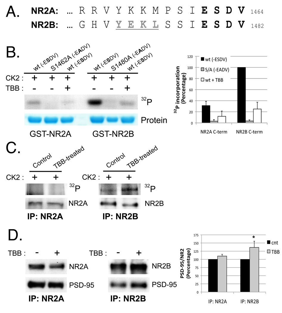
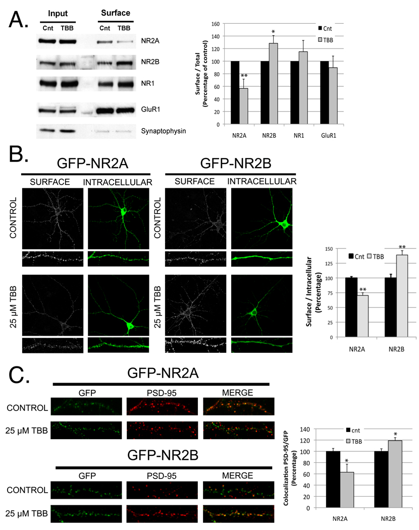
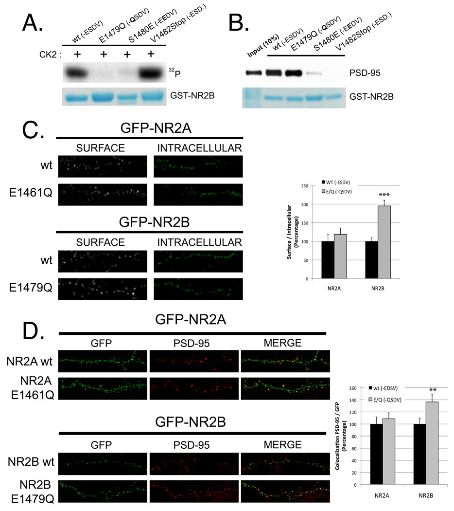
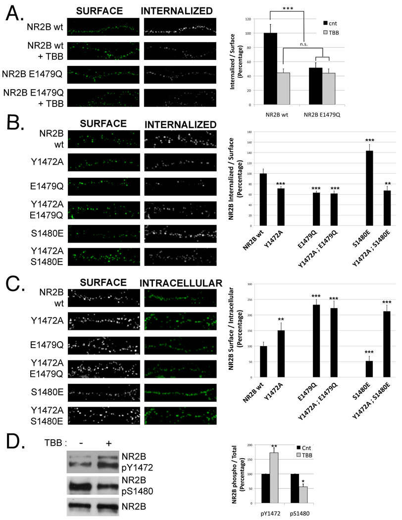
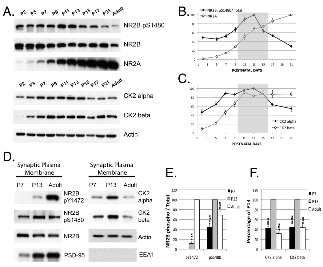
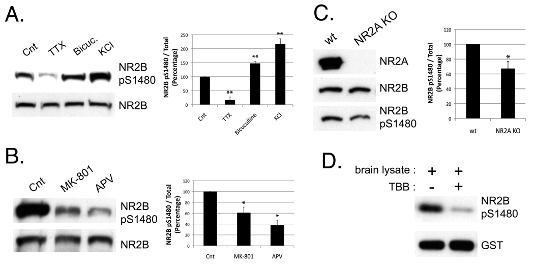
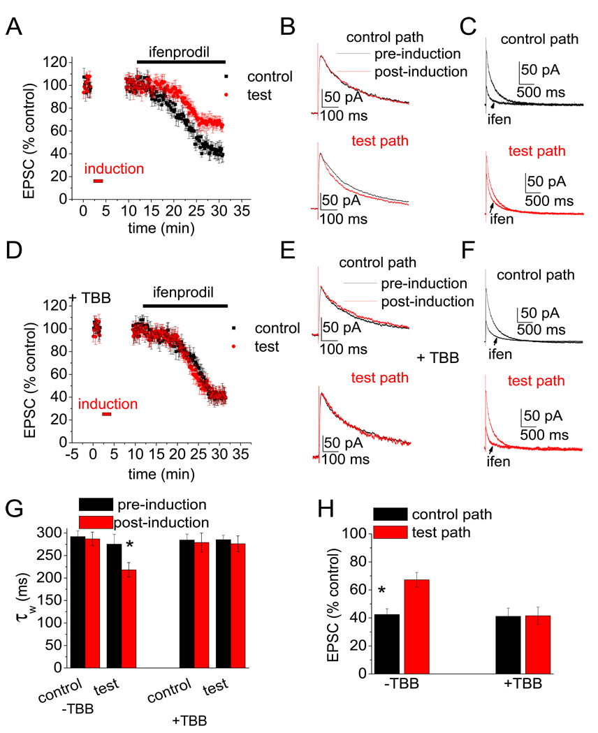
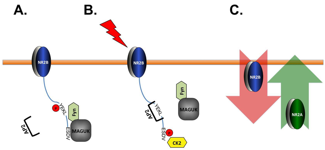
N-methyl-D-aspartate (NMDA) receptors (NMDARs) play a central role in development, synaptic plasticity, and neurological disease. NMDAR subunit composition defines their biophysical properties and downstream signaling. Casein kinase 2 (CK2) phosphorylates the NR2B subunit within its PDZ-binding domain; however, the consequences for NMDAR localization and function are unclear. Here we show that CK2 phosphorylation of NR2B regulates synaptic NR2B and NR2A in response to activity. We find that CK2 phosphorylates NR2B, but not NR2A, to drive NR2B-endocytosis and remove NR2B from synapses resulting in an increase in synaptic NR2A expression. During development there is an activity-dependent switch from NR2B to NR2A at cortical synapses. We observe an increase in CK2 expression and NR2B phosphorylation over this same critical period and show that the acute activity-dependent switch in NR2 subunit composition at developing hippocampal synapses requires CK2 activity. Thus, CK2 plays a central role in determining the NR2 subunit content of synaptic NMDARs.


Figures

Figure 1
Figure 1. CK2 phosphorylates NR2B much more efficiently than NR2A
A) Alignment of the extreme C-termini of rodent NR2A and NR2B. The PDZ binding domain (-ESDV) is shown in bold and the tyrosine-based endocytic motif in NR2B is underlined in grey. B) In vitro phosphorylation of the last 175 a.a. of NR2A (1289–1464) or NR2B (1307–1482) by CK2. GST-NR2A, GST-NR2A S1462A, GST-NR2B or GST-NR2B S1480A was incubated with CK2 and [γ-32P]ATP for 20 minutes at 30°C. When indicated, TBB (25 µM) was added to the sample. C) Cortical cultures (DIV10) were incubated with 25 µM TBB overnight to reduce endogenous phosphorylation of CK2 substrates. Cells were lysed and receptors recovered using specific NR2A or NR2B antibodies. Immunoprecipitates were subjected to an in vitro phosphorylation assay (as in main Figure 1B) with recombinant CK2 for 20 min at 30°C. n=3 D) HEK293 cells were co-transfected with PSD-95, NR1, and GFP-NR2A or GFP-NR2B. After treatment with TBB for 4 hours, cells were lysed in PBS with 1% TX-100. The amount of PSD-95 bound to NMDAR subunits was analyzed by co-immunoprecipitation with anti-NR2A or anti-NR2B antibodies. Graph represents means +/− SEM (n=4) *p<0.05
Figure 2
Figure 2. Inhibition of CK2 decreases surface and synaptic NR2A expression, but increases surface and synaptic NR2B
A) Cortical cultures (DIV 10) were treated overnight with 20 µM TBB or vehicle. Surface proteins were biotinylated and isolated with streptavidin-agarose beads as described in Experimental Procedures. Proteins were resolved by SDS-PAGE and blotted for NR2A, NR2B, NR1, GluR1 or synaptophysin (as control). Graph represents means +/− SEM *p < 0.05 **p < 0.01 (n=5) B) Hippocampal neurons expressing GFP-NR2A or GFP-NR2B were treated +/− TBB (25 µM). Surface receptors were labeled with anti-GFP antibody and Alexa-568 conjugated anti-rabbit secondary antibody (shown in white). After permeabilization, the internal pool of receptors was visualized by labeling with anti-GFP and Alexa-633 conjugated anti-rabbit secondary antibody (shown in green). n for NR2A (−/+ TBB) = 21, 16 ; n for NR2B (−/+ TBB) = 17, 22. Data represent means +/− SEM **p < 0.01. C) Colocalization of endogenous PSD-95 and NR2B or NR2A was evaluated in hippocampal neurons treated with 25 µM TBB, after transient transfection with GFP-NR2A or GFP-NR2B. n for NR2A(−/+ TBB) = 15, 18 ; n for NR2B (−/+ TBB) = 21, 28. Data represent means +/− SEM *p< 0.05. See also Figure S1
Figure 3
Figure 3. NR2B E1479Q, which is not phosphorylated by CK2, shows increased surface expression and synaptic localization compared to NR2B wt
A) In vitro CK2 phosphorylation (as described in Figure 1B) of the last 175 a.a. of NR2B attached to GST (wt, E1479Q, S1480E and V1482Stop) B) Pull-down experiments of GST-NR2B (wt, E1479Q, S1480E and V1482Stop). Beads were incubated with lysate of HEK293 cells expressing PSD-95 for 2 hours at 4°C. After washes, the recovered material was analyzed by immunoblotting with an anti-PSD-95 antibody. C) Surface expression analysis (as described in Figure 2B) was performed with hippocampal neurons expressing GFP-NR2A (wt or E1461Q) or GFP-NR2B (wt or E1479Q). Graph represents mean +/− SEM ***p<0.001. n for NR2A (wt, E/Q) =25, 21. n for NR2B (wt, E/Q) = 20, 26 D) Colocalization of endogenous PSD-95 with GFP-NR2A (wt or E1461Q) and GFP-NR2B (wt or E1479Q) analyzed at DIV14 as indicated in Figure 2C. Graph indicates mean +/− SEM. **p<0.01. n for NR2A (wt, E/Q) =17, 17. n for NR2B (wt, E/Q) = 16, 14.
Figure 4
Figure 4. Phosphorylation of NR2B S1480 increases endocytosis via a coordinated dephosphorylation of Y1472 in the YEKL endocytic domain
A) An endocytosis assay of hippocampal neurons transfected with GFP-NR2B (wt or E1479Q) was performed as described in Experimental Procedures. At DIV10, neurons were labeled with anti-GFP antibody, washed and returned to conditioned media (+/− 25 µM TBB) for 30 min at 37 °C to allow receptor internalization. Cells were fixed, and surface-expressed proteins were labeled with Alexa 568-conjugated secondary antibody (shown in green). After permeabilization with 0.25% TX-100, internalized receptors were labeled with Alexa 633-conjugated secondary antibody (shown as white). n for NR2B wt (+/− TBB) = 15, 25. n for NR2B E1479Q (+/− TBB) = 25, 21. Graph represents means +/− SEM ***p<0.001; n.s. denotes not significant differences. B) Endocytosis assay with NR2B constructs with mutations in the YEKL and/or PDZ-domain performed as in panel A. n= 31, 25, 29, 27, 15, 26. Graph represents means +/− SEM **p<0.01 ***p<0.001 C) Surface expression of NR2B constructs with mutations in the YEKL and/or PDZ-domain was analyzed as in Figure 2B. n= 38, 32, 22, 32, 17, 30. Graph represents means +/− SEM **p<0.01 ; ***p<0.001 D) Levels of NR2B phosphorylation (Y1472 and S1480) were analyzed in HEK293T cells transfected with PSD-95, NR1 and GFP-NR2B and incubated +/− 25 µM TBB for 4 hours. n=3. Graph represents means +/−SEM *p<0.05 ; ** p<0.01. See also Figure S2.
Figure 5
Figure 5. Phosphorylation of NR2B S1480 and total expression of CK2 increase during the second postnatal week. In addition, association of CK2 with synaptic plasma membranes is elevated at P13
A) Protein expression was analyzed in cortical synaptosomes from mice of different stages of development by immunoblotting. Graphs shown in panels B and C summarize the data of 6 experiments analyzing the level of NR2B Ser1480 phosphorylation and expression of NR2A and CK2 (alpha and beta subunits) throughout development (the grey area indicates the critical period of time for the NMDAR subunit switch). D) Synaptic plasma membranes (SPMs) were isolated from P7, P13 or adult animals, using a standard purification protocol (Hallet, 2008). The level of phosphorylated NR2B (Y1472 and S1480), CK2 (alpha and beta), EEA1 (as negative control) and actin (as loading control) present in the SPMs was analyzed by immunoblotting. E–F) Graph represents means +/− SEM ***p<0.001 n=6
Figure 6
Figure 6. Phosphorylation of NR2B on S1480 increases in response to NMDAR-activity and is regulated by NR2A expression
A) Cortical cultures (DIV10) were incubated for 8 hours with Tetrodoxin (TTX; 2 µM) or Bicuculline (Bicuc.; 40 µM) to block or stimulate neuronal activity respectively. Treatment with KCl (20 mM for 5 min) was used to induce neuronal depolarization. The level of phosphorylated NR2B was analyzed by immunoblotting after isolation of cellular membranes. The same membrane was re-blotted for NR2B or phospho-specific NR2B S1480 antibodies. Graph represents means +/− SEM **p<0.01 n=5 B) Cortical cultures were incubated overnight +/− NMDAR antagonists (100 µM APV ; 40 µM MK-801). Graph represents means +/− SEM *p<0.05 n=3 C) Cortical synaptosomes were isolated from P11 mice (wt or NR2A knock out) and analyzed by immunoblotting with the indicated antibodies. Graph represents means +/− SEM *p<0.05 n=4 D) GST-NR2B (last 175 a.a.) was phosphorylated in vitro using 10 µg of brain lysate as source of kinases. 25 µM TBB was added to the sample when indicated.
Figure 7
Figure 7. CK2 activity regulates the activity-dependent switch in the subunit composition of synaptic NMDA receptors
A) Pooled data (n = 10) for NMDA EPSC amplitude vs. time from control experiments showing the effect of LTP induction on ifenprodil block (red is test pathway to which the LTP induction protocol was applied; black is the control pathway; these colors are consistent throughout the figure). B) NMDA EPSCs from example control experiment showing the speeding of kinetics in test path after induction (lower panel). C) NMDA EPSCs from example control experiment showing the reduced block by ifenprodil (5 µM) in test path after induction (lower panel). D-F) As for control experiments (A–C), but in slices incubated with TBB (10 µM) for at least 2 hours (n = 7). G) Summary data of weighted decay time constant (control [−TBB] n = 10; +TBB n = 8; * indicates p < 0.05 between pre- and post-induction). H) Summary data of NMDA EPSC amplitude in ifenprodil (% of EPSC amplitude in absence of ifenprodil; control [−TBB] n = 10; +TBB n = 7; * indicates p < 0.05 between control and test pathways)
Figure 8
Figure 8. Model of CK2 regulation of synaptic NMDARs
A) Early in development, the association of NR2B with MAGUKs stabilizes NR2B at synaptic membranes via phosphorylation of Y1472 by Fyn. Phosphorylation of the Y1472 within the tyrosine-based endocytic motif blocks AP-2 binding. B) During the critical period, NMDAR activity induces NR2B S1480 phosphorylation by CK2, which results in the disruption of NR2B association with MAGUKs. NR2B Y1472 is now dephosphorylated and AP-2 can bind to the YEKL motif and promote NR2B endocytosis. C) NR2A expression increases and NR2A-containing receptors replace NR2B-containing NMDARs at synaptic sites.

Similar articles

Cited by

References

    1. Aksenova MV, Burbaeva GS, Kandror KV, Kapkov DV, Stepanov AS. The decreased level of casein kinase 2 in brain cortex of schizophrenic and Alzheimer's disease patients. FEBS letters. 1991;279:55–57. - PubMed
    1. Al-Hallaq RA, Conrads TP, Veenstra TD, Wenthold RJ. NMDA di-heteromeric receptor populations and associated proteins in rat hippocampus. J Neurosci. 2007;27:8334–8343. - PMC - PubMed
    1. Allende JE, Allende CC. Protein kinases. 4. Protein kinase CK2: an enzyme with multiple substrates and a puzzling regulation. Faseb J. 1995;9:313–323. - PubMed
    1. Alvestad RM, Grosshans DR, Coultrap SJ, Nakazawa T, Yamamoto T, Browning MD. Tyrosine dephosphorylation and ethanol inhibition of N-Methyl-D-aspartate receptor function. The Journal of biological chemistry. 2003;278:11020–11025. - PubMed
    1. Barria A, Malinow R. Subunit-specific NMDA receptor trafficking to synapses. Neuron. 2002;35:345–353. - PubMed

Publication types

MeSH terms

Substances

LinkOut - more resources