. 2010 Sep 23;67(6):1009-20.
doi: 10.1016/j.neuron.2010.08.014.

Ultrastructural analysis of hippocampal neuropil from the connectomics perspective

Affiliations

Ultrastructural analysis of hippocampal neuropil from the connectomics perspective

Yuriy Mishchenko et al. Neuron. .

Abstract

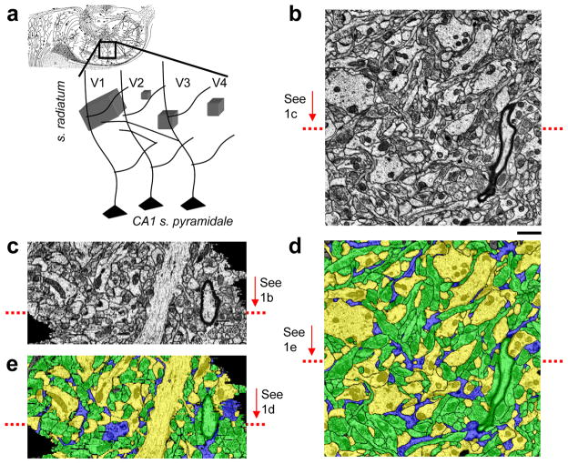
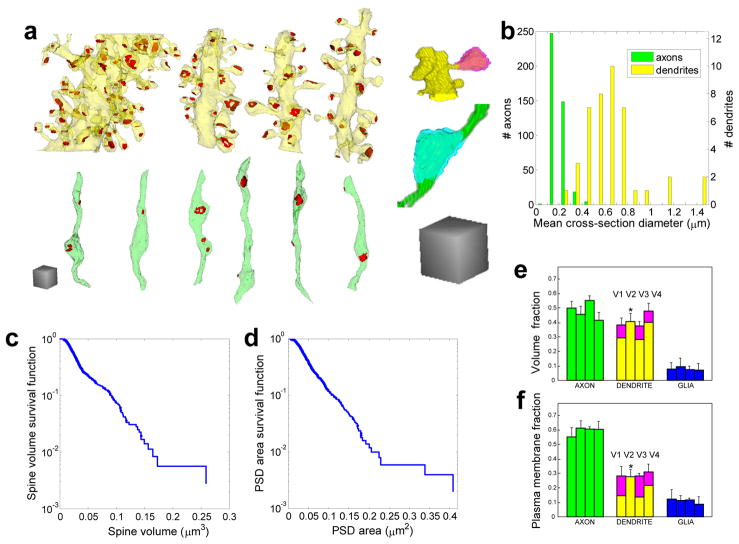
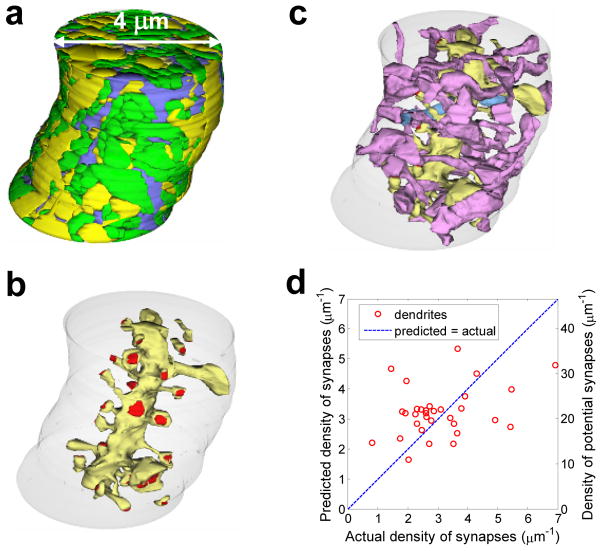
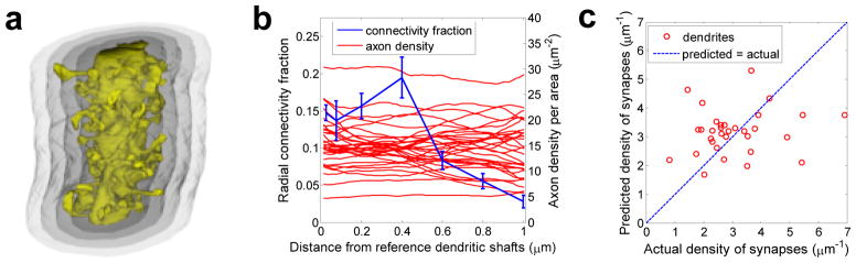
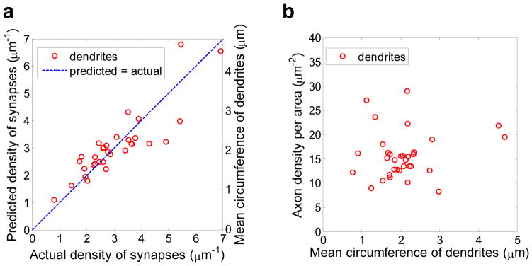
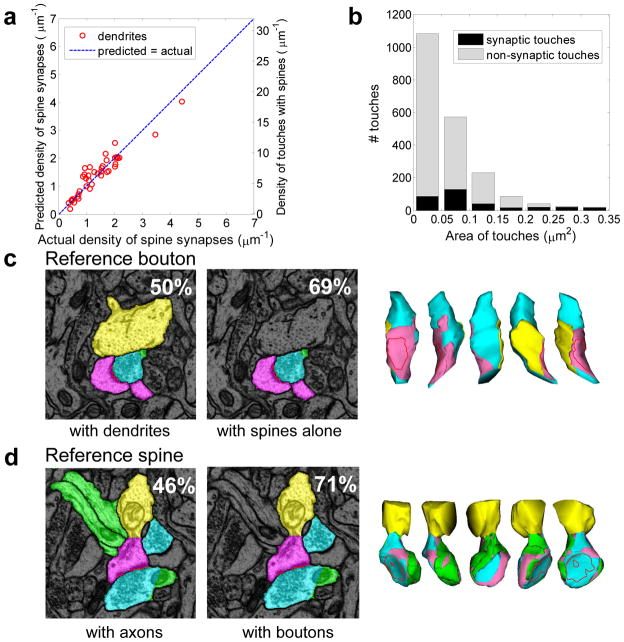
Complete reconstructions of vertebrate neuronal circuits on the synaptic level require new approaches. Here, serial section transmission electron microscopy was automated to densely reconstruct four volumes, totaling 670 μm(3), from the rat hippocampus as proving grounds to determine when axo-dendritic proximities predict synapses. First, in contrast with Peters' rule, the density of axons within reach of dendritic spines did not predict synaptic density along dendrites because the fraction of axons making synapses was variable. Second, an axo-dendritic touch did not predict a synapse; nevertheless, the density of synapses along a hippocampal dendrite appeared to be a universal fraction, 0.2, of the density of touches. Finally, the largest touch between an axonal bouton and spine indicated the site of actual synapses with about 80% precision but would miss about half of all synapses. Thus, it will be difficult to predict synaptic connectivity using data sets missing ultrastructural details that distinguish between axo-dendritic touches and bona fide synapses.


Figures

Figure 1
Figure 1
Reconstructed volumes. a) Location of the four volumes (V1–4) relative to CA1 pyramidal neuron dendrites in the hippocampus; b) Typical ssTEM micrograph of the hippocampus neuropil from V1; c) V1 re-sectioned orthogonal to the cutting plane at the location indicated by the red arrow in b. Note that the stack is well aligned and the ultrastructure is visible despite lower z-resolution; d) Electron micrograph from b after automated segmentation and proofreading colored according to the object class: axons - green, dendrites - yellow, and glia processes - blue; e) Segmented re-section from c. Scale bar:1μm (b–e)
Figure 2
Figure 2
Shapes and dimensions of various objects in the neuropil. a) Three-dimensional reconstruction of representative objects in V3: dendrites (yellow), axons (green), post-synaptic densities (PSDs) (red), spine (pink), and bouton (cyan); b) Distribution of the effective axonal and dendritic cross-section diameters in V1 and V3; c) Survival function of spine volume, i.e. a fraction of spines whose volume is less than a given value; d) survival function of the PSD area. Only spines and PSDs completely contained within V1 were included in c) and d); e) Distribution of volume among different object classes in the four volumes; f) Distribution of plasma membrane surface area among different object classes. Scale cubes in a) are 1 μm on the side; bars in e) and f) are arranged sequentially V1 → V4 in each object class (axon, dendrite, glia); * in e) and f) - calculations of the volume of spine heads and other analysis were not performed for V2 given its small size.
Figure 3
Figure 3
Comparison of actual density of synapses along individual dendrites in V1 and V3 and predictions based on maximum reach connectivity fraction. a) Manual reconstruction of cylinder centered on the central oblique dendrite coursing through V1 and containing axons (green), dendrites (yellow), and glia (blue). Double arrowed line indicates the diameter of the cylinder. b) Central oblique dendrite (yellow) and its associated synapses (red) located on dendritic spines. The boundary of the smallest neuropil cylinder that contained the selected oblique dendrite and all of its spines is illustrated in light gray. c) Subpopulation of axons (purple, to distinguish from all green axons in a)) that formed synapses with the central oblique dendrite (yellow). Of these 28 axons, 27 made just one synapse and 1 made 2 synapses (light blue axon) on this dendrite. d) Plot of the actual density of synapses for dendrites in V1 and V3 vs. the density of synapses predicted by multiplying the mean maximum-reach connectivity fraction by the local density of potential synapses. This method is a weak predictor (r2 ≈ 0.12).
Figure 4
Figure 4
Comparison of actual density of synapses along individual dendrites in V1 and V3 and predictions based on the distance dependent connectivity fraction. a) 3D illustration of one dendritic segment and four radial shells, each following the surface outline of the dendritic shaft after the spines had been truncated. b) Dependence of the mean connectivity fraction and axonal density on the distance from the surface of the dendritic shaft. c) Plot of the actual density of synapses along dendrites in V1 and V3 vs. the density of synapses predicted by convolving the mean distance dependent connectivity fraction (blue line in b) with the local axon density (red lines in b). This method is a weak predictor (r2 ≈ 0.02).
Figure 5
Figure 5
Relationship between dendritic caliber and the density of actual and potential synapses. a) Plot of the actual density of synapses vs. the density of synapses predicted by multiplying the dendritic circumference by the common coefficient. Dendritic caliber is a strong predictor of actual density of synapses along a dendrite (r2 ≈ 0.75). b) Density of available axons (per unit length of dendrite per unit distance from a dendrite) does not correlate with the dendritic caliber (r2 ≈ 0.02).
Figure 6
Figure 6
Relationship between synaptic and non-synaptic axo-dendritic touches in V1. a) Density of spine synapses along a dendrite is proportional to the density of spine touches with axons (r2 ≈ 0.88); b) Area distributions of synaptic and non-synaptic touches overlap significantly. c) Reference bouton whose largest touch with a spine corresponds to a synapse. Left: section containing the reference bouton (cyan) with touching dendrite (yellow) and spine (pink). Percentage of boutons with largest dendritic touch corresponding to a synapse is shown. Center: Reference bouton (cyan) and touching spine (pink) form a synapse. Percentage of boutons with largest spine touch corresponding to a synapse is shown. Right: 3D views of the reference bouton colored according to the type of touching object. Visible blue areas are where other axons touched this bouton. d) Reference spine whose largest touch with a bouton corresponds to a synapse. Left: section containing the reference spine (pink) with touching axons (green) and boutons (cyan). Percentage of spines with largest axonal touch corresponding to a synapse is shown. Center: Reference spine (pink) and touching boutons (cyan). Percentage of spines with largest bouton touch corresponding to a synapse. Right: 3D views of the reference spine surface colored according to the type of touching object. Red dotted line: position of the synapse.

References

    1. Anderson JR, Jones BW, et al. A computational framework for ultrastructural mapping of neural circuitry. PLoS Biol. 2009;7(3):e1000074. - PMC - PubMed
    1. Benjamini Y, Hochberg Y. Controlling the False Discovery Rate - a Practical and Powerful Approach to Multiple Testing. Journal of the Royal Statistical Society Series B-Methodological. 1995;57(1):289–300.
    1. Benjamini Y, Yekutieli D. The control of the false discovery rate in multiple testing under dependency. Annals of Statistics. 2001;29(4):1165–1188.
    1. Biederer T, Sara Y, et al. SynCAM, a synaptic adhesion molecule that drives synapse assembly. Science. 2002;297(5586):1525–1531. - PubMed
    1. Binzegger T, Douglas RJ, et al. A quantitative map of the circuit of cat primary visual cortex. J Neurosci. 2004;24(39):8441–8453. - PMC - PubMed

Publication types

LinkOut - more resources