Biological synthesis of tooth enamel instructed by an artificial matrix
- PMID: 20869764
- PMCID: PMC2956865
- DOI: 10.1016/j.biomaterials.2010.08.013
Biological synthesis of tooth enamel instructed by an artificial matrix
Abstract
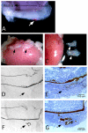
The regenerative capability of enamel, the hardest tissue in the vertebrate body, is fundamentally limited due to cell apoptosis following maturation of the tissue. Synthetic strategies to promote enamel formation have the potential to repair damage, increase the longevity of teeth and improve the understanding of the events leading to tissue formation. Using a self-assembling bioactive matrix, we demonstrate the ability to induce ectopic formation of enamel at chosen sites adjacent to a mouse incisor cultured in vivo under the kidney capsule. The resulting material reveals the highly organized, hierarchical structure of hydroxyapatite crystallites similar to native enamel. This artificially triggered formation of organized mineral demonstrates a pathway for developing cell fabricated materials for treatment of dental caries, the most ubiquitous disease in man. Additionally, the artificial matrix provides a unique tool to probe cellular mechanisms involved in tissue formation further enabling the development of tooth organ replacements.
Copyright © 2010 Elsevier Ltd. All rights reserved.
Figures


Similar articles
-
Biomimetic Remineralization of Carious Lesions by Self-Assembling Peptide.J Dent Res. 2017 Jul;96(7):790-797. doi: 10.1177/0022034517698419. Epub 2017 Mar 27. J Dent Res. 2017. PMID: 28346861
-
Evaluation of the effect of self-assembling peptide and fluoride varnish, alone or in combination with laser irradiation, on artificial enamel caries: a SEM/EDS and Micro-CT study.Clin Oral Investig. 2024 Aug 28;28(9):503. doi: 10.1007/s00784-024-05901-1. Clin Oral Investig. 2024. PMID: 39196467
-
Protein Interaction between Ameloblastin and Proteasome Subunit α Type 3 Can Facilitate Redistribution of Ameloblastin Domains within Forming Enamel.J Biol Chem. 2015 Aug 21;290(34):20661-20673. doi: 10.1074/jbc.M115.640185. Epub 2015 Jun 12. J Biol Chem. 2015. PMID: 26070558 Free PMC article.
-
Enamel maturation.Ciba Found Symp. 1997;205:156-70; discussion 170-4. doi: 10.1002/9780470515303.ch11. Ciba Found Symp. 1997. PMID: 9189623 Review.
-
The structural biology of the developing dental enamel matrix.J Struct Biol. 1999 Jun 30;126(3):270-99. doi: 10.1006/jsbi.1999.4130. J Struct Biol. 1999. PMID: 10441532 Review.
Cited by
-
Elastin-like peptide amphiphiles form nanofibers with tunable length.Biomacromolecules. 2012 Sep 10;13(9):2645-54. doi: 10.1021/bm300472y. Epub 2012 Aug 21. Biomacromolecules. 2012. PMID: 22849577 Free PMC article.
-
Design of materials with supramolecular polymers.Prog Polym Sci. 2020 Dec;111:101310. doi: 10.1016/j.progpolymsci.2020.101310. Epub 2020 Oct 15. Prog Polym Sci. 2020. PMID: 33082608 Free PMC article. Review.
-
Supramolecular biomaterials.Nat Mater. 2016 Jan;15(1):13-26. doi: 10.1038/nmat4474. Nat Mater. 2016. PMID: 26681596 Review.
-
Nanotechnology in dentistry: prevention, diagnosis, and therapy.Int J Nanomedicine. 2015 Oct 8;10:6371-94. doi: 10.2147/IJN.S86033. eCollection 2015. Int J Nanomedicine. 2015. PMID: 26504385 Free PMC article. Review.
-
Harnessing biomolecules for bioinspired dental biomaterials.J Mater Chem B. 2020 Oct 14;8(38):8713-8747. doi: 10.1039/d0tb01456g. Epub 2020 Aug 4. J Mater Chem B. 2020. PMID: 32747882 Free PMC article. Review.
References
-
- Taubman MA, Nash DA. The scientific and public-health imperative for a vaccine against dental caries. Nat Rev Immunol. 2006;6:555–63. - PubMed
-
- Robinson C, Shore RC, Brookes SJ, Strafford S, Wood SR, Kirkham J. The chemistry of enamel caries. Crit Rev Oral Biol Med. 2000;11:481–95. - PubMed
-
- Caufield PW, Li Y, Dasanayake A. Dental caries: an infectious and transmissible disease. Compend Contin Educ Dent. 2005;26:10–6. - PubMed
-
- Hu B, Nadiri A, Kuchler-Bopp S, Perrin-Schmitt F, Peters H, Lesot H. Tissue Engineering of Tooth Crown, Root, and Periodontium. Tissue Eng. 2006;12:2069–75. - PubMed
-
- Mitsiadis TA, Graf D. Cell fate determination during tooth development and regeneration. Birth Defects Res C Embryo Today. 2009;87:199–211. - PubMed
Publication types
MeSH terms
Substances
Grants and funding
LinkOut - more resources
Full Text Sources
Other Literature Sources

