

Monoamine oxidases (MAO) in the pathogenesis of heart failure and ischemia/reperfusion injury
- PMID: 20869994
- PMCID: PMC3030628
- DOI: 10.1016/j.bbamcr.2010.09.010
Monoamine oxidases (MAO) in the pathogenesis of heart failure and ischemia/reperfusion injury
Abstract
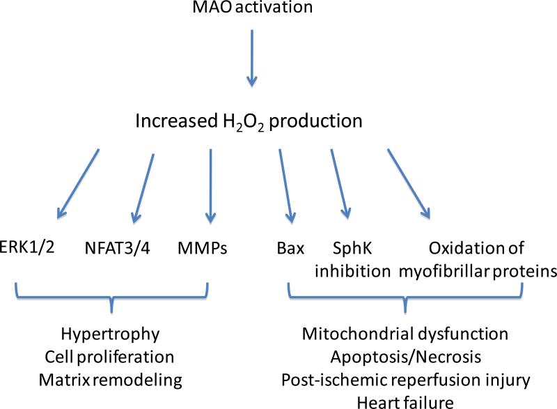
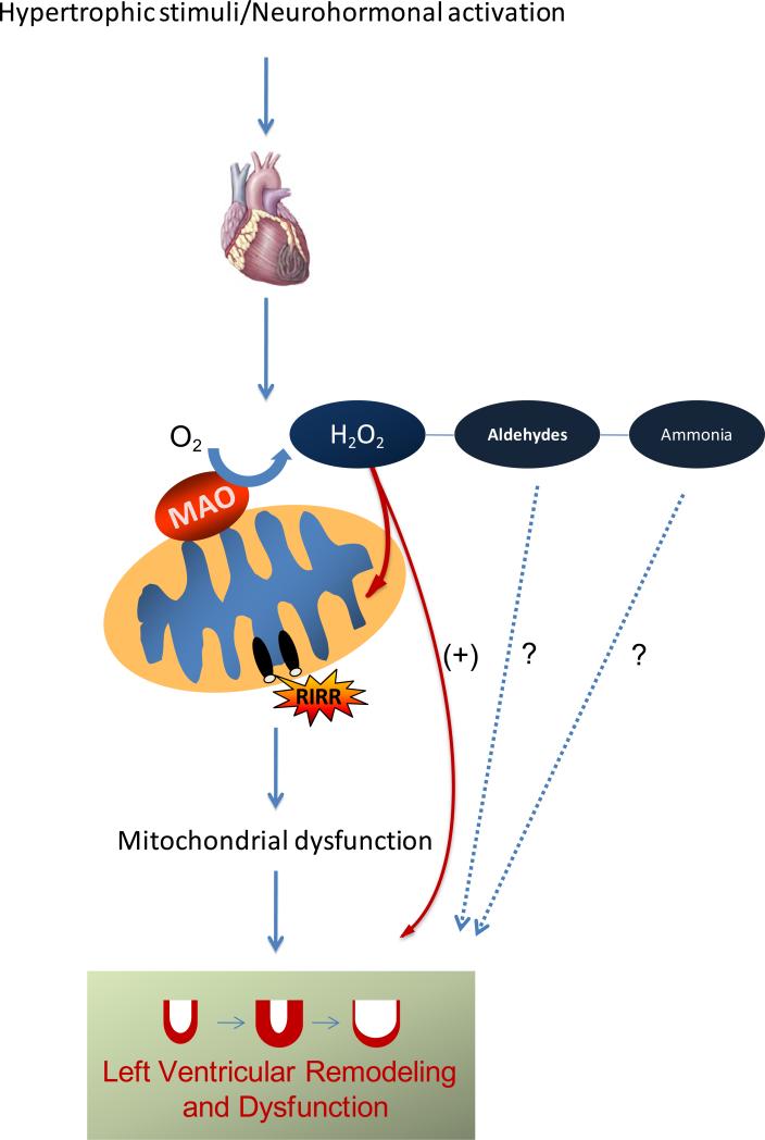
Recent evidence highlights monoamine oxidases (MAO) as another prominent source of oxidative stress. MAO are a class of enzymes located in the outer mitochondrial membrane, deputed to the oxidative breakdown of key neurotransmitters such as norepinephrine, epinephrine and dopamine, and in the process generate H(2)O(2). All these monoamines are endowed with potent modulatory effects on myocardial function. Thus, when the heart is subjected to chronic neuro-hormonal and/or peripheral hemodynamic stress, the abundance of circulating/tissue monoamines can make MAO-derived H(2)O(2) production particularly prominent. This is the case of acute cardiac damage due to ischemia/reperfusion injury or, on a more chronic stand, of the transition from compensated hypertrophy to overt ventricular dilation/pump failure. Here, we will first briefly discuss mitochondrial status and contribution to acute and chronic cardiac disorders. We will illustrate possible mechanisms by which MAO activity affects cardiac biology and function, along with a discussion as to their role as a prominent source of reactive oxygen species. Finally, we will speculate on why MAO inhibition might have a therapeutic value for treating cardiac affections of ischemic and non-ischemic origin. This article is part of a Special Issue entitled: Mitochondria and Cardioprotection.
Copyright © 2010 Elsevier B.V. All rights reserved.
Figures
References
-
- Taegtmeyer H. Energy metabolism of the heart: from basic concepts to clinical applications. Curr. Probl. Cardiol. 1994;19:59–113. - PubMed
-
- Stanley WC, Chandler MP. Energy metabolism in the normal and failing heart: potential for therapeutic interventions. Heart Fail. Rev. 2002;7:115–130. - PubMed
-
- BING RJ, SIEGEL A, UNGAR I, GILBERT M. Metabolism of the human heart. II. Studies on fat, ketone and amino acid metabolism. Am. J. Med. 1954;16:504–515. - PubMed
-
- Halestrap AP, Kerr PM, Javadov S, Woodfield KY. Elucidating the molecular mechanism of the permeability transition pore and its role in reperfusion injury of the heart. Biochim. Biophys. Acta. 1998;1366:79–94. - PubMed
-
- Jennings RB, Ganote CE. Mitochondrial structure and function in acute myocardial ischemic injury. Circ. Res. 1976;38:I80–I91. - PubMed
Publication types
MeSH terms
Substances
Grants and funding
LinkOut - more resources
Full Text Sources
Medical
Molecular Biology Databases
