. 2010 Dec 15;49(12):1872-80.
doi: 10.1016/j.freeradbiomed.2010.09.012. Epub 2010 Sep 24.

RNA interference against CFTR affects HL60-derived neutrophil microbicidal function

Affiliations

RNA interference against CFTR affects HL60-derived neutrophil microbicidal function

Ryan W Bonvillain et al. Free Radic Biol Med. .

Abstract

Biosynthesis of hypochlorous acid, a potent antimicrobial oxidant, in phagosomes is one of the chief mechanisms employed by polymorphonuclear neutrophils to combat infections. This reaction, catalyzed by myeloperoxidase, requires chloride anion (Cl(-)) as a substrate. Thus, Cl(-) availability is a rate-limiting factor that affects neutrophil microbicidal function. Our previous research demonstrated that defective CFTR, a cAMP-activated chloride channel, present in cystic fibrosis (CF) patients leads to deficient chloride transport to neutrophil phagosomes and impaired bacterial killing. To confirm this finding, here we used RNA interference against this chloride channel to abate CFTR expression in the neutrophil-like cells derived from HL60 cells, a promyelocytic leukemia cell line, with dimethyl sulfoxide. The resultant CFTR deficiency in the phagocytes compromised their bactericidal capability, thereby recapitulating the phenotype seen in CF patient cells. The results provide further evidence suggesting that CFTR plays an important role in phagocytic host defense.


Figures

Figure 1
Figure 1. CFTR expression in HL60 cells and their derived neutrophil-like cells
A) Transcription of CFTR – Reverse transcription PCR using exon-specific primers produced cDNA-specific amplification of CFTR fragment, confirming that PMNs and HL60 cells express CFTR at the mRNA level. The reverse transcriptase-free [RT(−)] and primer-free [Primer(−)] controls showed no specific amplification. B-F) Subcellular localization of CFTR – DMSO-differentiated HL60 cells were fed serum-opsonized zymosan at a ratio of 5:1 (zymosan to neutrophil) and were subsequently stained with either mouse IgG2a isotype or mouse anti-CFTR 24-1 primary antibody. B & D) Relief contrast images show engulfment of zymosan particles by the differentiated neutrophils. C & E) Fluorescence microscopy shows that CFTR-positive granules distribute around the phagocytosed particles (arrows). F) An enlarged image of CFTR staining shows the intracellular punctate staining pattern and potential granule fusion (arrows).
Figure 2
Figure 2. Small interfering RNA to suppress CFTR expression
A) Structures of the transgene constructs – The short hairpin CFTR sequence (shCFTR) and the scrambled control sequence (shControl) were under the transcriptional control of the mouse U6 promoter. The eGFP reporter gene is an independent expression cassette driven by the cytomegalovirus (CMV) promoter. Micrographs by relief contrast and fluorescent microscopy of the parental HL60 (B & C), HL60-shControl (E & F) and HL60-shCFTR (H & I) cells showed expression of eGFP in the transduced cells which was confirmed by flow cytometry analysis of each cell subtype (D, G, & J, respectively).
Figure 3
Figure 3. CFTR expression in the control and CFTR-knockdown HL60-derived neutrophils
A) Quantitative RT-PCR of CFTR transcripts as normalized to GAPDH – HL60-shCFTR derived cells had 84.2% less CFTR expression than that of the HL60-shControl cells, confirming CFTR knockdown by the siRNA. B) Immunoprecipitation and in vitro phosphorylation to assess CFTR protein expression – an equal number of DMSO-differentiated HL60-shControl and HL60-shCFTR neutrophil-like cells were lysed and the cell lysates were immunoprecipitated for CFTR protein. In vitro phosphorylation with radioactive [γ32P]-ATP was performed to label CFTR followed by SDS-PAGE. Autoradiography displays CFTR-specific band at ~180 KDa (top panel). Ponceau S staining of a membrane transferred with the total cell lysate proteins after gel electrophoresis was performed. The β-actin band at ~42 KDa is specifically displayed to shown an equal protein application to the immunoprecipitation (bottom panel). C-H) Flow cytometry analysis of CFTR expression on phagosomes – Both HL60-shControl- and HL60-shCFTR-derived neutrophils were allowed to phagocytose serum-opsonized 1-μm latex beads. After phagocytosis, the cells were homogenized by nitrogen cavitation. Antibody staining and flow cytometry were performed on the cell lysates by adding a Cy3-conjugated antibody against human lysosome-associated membrane protein-1 (LAMP-1) and a mouse anti-human CFTR antibody. C & F) The forward scatter (FSC) and side scatter (SSC) parameters of the stained cell lysates were adjusted to gate on the 1-μm bead population. D & G) The 1-μm beads were analyzed for LAMP-1 and CFTR expression. The dual-positive population represents phagolysosome membranes containing both LAMP-1 and CFTR. E & H) The dual-positive beads were further analyzed for CFTR expression levels. The shaded lines represent the CFTR antibody staining. The black lines represent the control isotype antibody staining.
Figure 4
Figure 4. Differentiation and phagocytosis of the DMSO-differentiated HL60-shControl and HL60-shCFTR cells
A) Differentiation of the differentiated cells – Both control and CFTR-knockdown cell lines were differentiated into neutrophil-like cells by exposure to 1.25% DMSO for 5 days and stained with Wright-Giemsa stain. Differentiation was classified based on the degree of nuclear polymorphism as follows: round nuclei – promyelocytes (undifferentiated), nuclei with apparent invaginations – metamyelocytes, elongated nuclei with distinct banding – banded neutrophils, polymorphic nuclei – segmented neutrophils. Percentages of each cell type in the total cell populations were calculated and compared between cell lines by Student’s t-test. There was no statistical difference in the proportion of the differentiated cells in each culture between the control and CFTR-knockdown cells. B) Phagocytosis by the differentiated cells – Differentiated cultures of HL60-shControl and HL60-shCFTR cells were fed serum-opsonized zymosan particles at a ratio of ~5-10:1 (particles to cells) for 15 minutes at 37°C. After phagocytosis, the cells were fixed and mounted onto glass slides by cytospin. The number of particles per phagocytic cell was determined microscopically and compared between control and CFTR-knockdown groups by Student’s t-test (P = 0.15, n = 3).
Figure 5
Figure 5. Hypochlorous acid (HOCl) production by control and CFTR-knockdown HL60-derived neutrophils
Both DMSO-differentiated HL60-shControl and HL60-shCFTR cells produced abundant HOCl when stimulated with PMA and supplied with physiological chloride in the extracellular media. The MPO inhibitor 4-aminobenzoic acid hydrazide (ABAH) and the NADPH oxidase inhibitor diphenylene iodonium (DPI) diminish the extracellular production of HOCl in this system. There is little HOCl produced when the cells are incubated in a chloride-free medium (NaGluconate Ringer’s buffer, 0 mM Cl). Throughout, there was no statistical difference in extracellular HOCl production between the control and CFTR-knockdown cell lines suggesting that the oxidant production machinery is unaffected by the CFTR knockdown genetic manipulation.
Figure 6
Figure 6. Killing of Pseudomonas aeruginosa by neutrophil-like cells derived from HL60-shControl and HL60-shCFTR cells
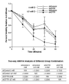
Percent viability (relative to time 0) of the phagocytosed bacteria by the control (shControl) or the knockdown (shCFTR) cells was plotted against time (Minutes). The graph represents 3 independent experiments. Two-way ANOVA analyses of the different group combinations were performed and the results shown below. In the absence of chloride, neutrophils differentiated from the control and knockdown cells had a low bacterial killing efficiency and showed no statistical difference. However, in the presence of 127 mM chloride, the control cells, but not the knockdown cells, showed an increased bactericidal activity with a statistically significant difference (p<0.0001, n=3). Therefore, the loss of CFTR in the knockdown cells impaired the chloride-dependent microbicidal capacity of the cells.
Figure 7
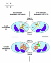
Figure 7. Proposed model to explain CFTR involvement in phagocytic microbicidal function
When intracellular and extracellular chloride was depleted (Panels A & C), the phagosomes of both control HL60-neutrophils (ShControl) and CFTR Knockdown (ShCFTR) HL60-neutrophils acquired little chloride. Thus, HOCl production was affected for both cells, showing no bacterial killing difference. However, when extracellular chloride was supplied after phagocytosis, only the control HL60-neutrophils (ShControl) (Panel B), but not the CFTR Knockdown (ShCFTR) HL60-neutrophils (Panel D) provided significant chloride to the phagosomes to generate HOCl, thus showing greater bacterial killing than the knockdown cells.

Similar articles

Cited by

References

    1. Klebanoff S. Myeloperoxidase: friend and foe. J Leukocyte Biol. 2005 May;77:598–625. - PubMed
    1. Nauseef W. How human neutrophils kill and degrade microbes: an integrated view. Immunol Rev. 2007;219:88–102. - PubMed
    1. Foote CS, Goyne TE, Lehrer RI. Assessment of chlorination by human neutrophils. Nature. 1983;301(5902):715–6. - PubMed
    1. Winterbourn CC, et al. Modeling the reactions of superoxide and myeloperoxidase in the neutrophil phagosome: implications for microbial killing. J Biol Chem. 2006;281(52):39860–9. - PubMed
    1. Murphy R, DeCoursey TE. Charge compensation during the phagocyte respiratory burst. Biochim Biophys Acta. 2006;1757(8):996–1011. - PubMed

Publication types

MeSH terms