Review
. 2011 Jan;21(1):47-56.
doi: 10.1016/j.tcb.2010.08.015. Epub 2010 Oct 1.

Forcing form and function: biomechanical regulation of tumor evolution

Affiliations
Review

Forcing form and function: biomechanical regulation of tumor evolution

Hongmei Yu et al. Trends Cell Biol. 2011 Jan.

Abstract

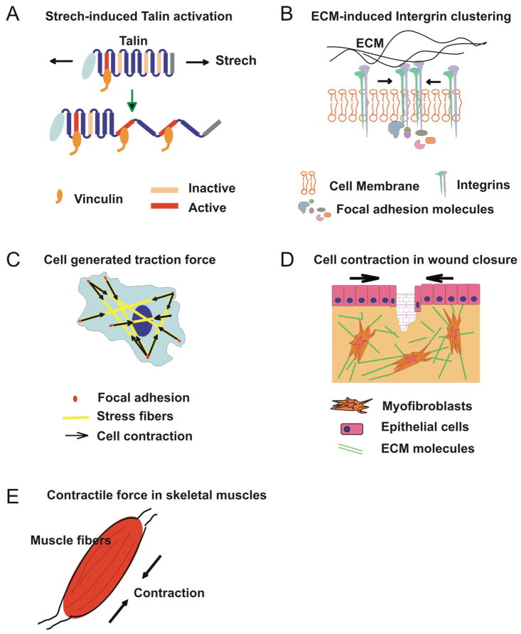
Cancer cells exist in a constantly evolving tissue microenvironment of diverse cell types within a proteinaceous extracellular matrix. As tumors evolve, the physical forces within this complex microenvironment change, with pleiotropic effects on both cell- and tissue-level behaviors. Recent work suggests that these biomechanical factors direct tissue development and modulate tissue homeostasis, and, when altered, crucially influence tumor evolution. In this review, we discuss the biomechanical regulation of cell and tissue homeostasis from the molecular, cellular and tissue levels, including how modifications of this physical dialogue could contribute to cancer etiology. Because of the broad impact of biomechanical factors on cell and tissue functions, an understanding of tumor evolution from the biomechanical perspective should improve risk assessment, clinical diagnosis and the efficacy of cancer treatment.


Figures

Figure 1
Figure 1. Dynamic and reciprocal conversation between matrix stiffness and cell tension
Non-malignant cells can respond to exogenous mechanical forces and matrix stiffness by enhancing focal complex maturation, resulting in Rho and ROCK activation, MLC phosphorylation and actomyosin contraction. Cell-generated forces from increased myosin contraction feed into focal adhesion maturation to adjust focal adhesion size and contract the ECM until the exogenous forces are balanced and the elastic modulus of cells are “tuned” to reflect the ECM stiffness. This mechano-signaling circuit is crucial in the dynamic and reciprocal conversation between exogenous and cell-generated mechanical factors. (ROCK, Rho-associated kinase; MLC, myosin light chain.) (Adapted with permission from [20].)
Figure 2
Figure 2. Examples of mechanical behaviors at different levels of biological systems
A) At the single-molecule level, mechanical stretching of talin rods exposes the cryptic binding sites for vinculin, which then activates downstream biochemical signaling pathways important in cell signaling, adhesion, and migration. B) At the multimolecule-level, increased matrix stiffness enhances integrin clustering, promotes large focal adhesions and activates downstream signaling cascades. C) At the single-cell level, cells can generate traction forces via actin polymerization (e.g. stress fibers) and actomyosin contraction between focal adhesions. D) At the tissue level, myofibroblasts differentiated from fibroblasts at wound sites exert contractile forces on the surrounding ECM and rearrange the ECM to close wounds. E) At the organ level, a muscle that contains multiple muscle fibers surrounded by connective tissues and sheaths can contract synchronously, generating tension and muscle motion upon the reception of signals from motor neurons.
Figure 3
Figure 3. Tumor progression is associated with continuous alterations in tissue and cell mechanics
A) In hyperplastic lesions, cells gradually lose polarity and grow into the luminal space. The myoepithelial cell layer and the basement membrane (BM) remain intact and the surrounding ECM is compliant. B) In carcinoma in situ lesions, cell polarity is lost and the lumen is filled by proliferating cells. This volume expansion and resistance from the BM and interstitial ECM lead to increased forces between tumor cells and the stromal matrix. Simultaneously, ECM components are abnormally deposited and remodeled, which results in increased ECM and tissue stiffness, and in turn, cell-generated tension. C) In invasive lesions, tumor cells break down the BM and invade into the interstitial ECM. The reciprocal forces between tumor cells and the ECM continuously increase. Abnormal deposition and remodeling of ECM collagen further increase ECM and tissue stiffness. Tumor cells generate greater tension in response to this increased mechanical stimulation. As tumor cells invade through the BM and ECM, they experience a range of different forces from the dense ECM network. These external forces together with genetic and epigenetic events can change the contractility and viscoelasticity of tumor cells. D) During intravasation and extravasation, tumor cells experience various forces including shear forces exerted by the ECM, blood flow and neighboring cells, which facilitate their transport and attachment to the endothelium. E) and F) Cancer cells often metastasize to different organs, which can have very different microenvironmental and mechanical properties (e.g. E. soft tissue lung, F. stiff tissue bone). The mechanics of remote tissues and cancer cells may regulate cell dormancy, proliferation and differentiation in these organs.
Figure 4
Figure 4. Increased collagen cross-linking and matrix stiffness modify the context of signaling and promote invasion of oncogene-transformed pre-malignant mammary cells
A) MCF10A cells (non-malignant mammary epithelial cells) expressing either a drug-activated ErbB2/NGFR (NGFR, neural growth factor receptor) chimera or a tetracycline-inducible ErbB2 construct form polarized, growth-arrested colonies in soft collagen/rBM gels (bottom left). Stiffening the collagen gel (by adding ribose to cross-link the collagen) or activating ErbB2 signaling increases cell proliferation but fails to drive cell invasion (bottom right and top left). MCF10A colonies start invading into collagen gels only when the collagen gels are stiffened and ErbB2 signaling is activated. (top right) (Bar 20 m;-catenin, green; 4 integrin, red; DAPI, blue.) Second harmonic generation images show the collagen alignment and bundling around the colonies (insert). B) Schematic presentation of the cooperation between matrix stiffening and ErbB2 signaling in driving the invasive phenotype. Increased collagen cross-linking stiffens the ECM, which drives integrin clustering and promotes focal adhesion assembly, thereby activating PI3K and potentiating ErbB2/PI3K/Akt signaling. (Adapted with permission from [40].)

References

    1. Paszek MJ, Weaver VM. The tension mounts: mechanics meets morphogenesis and malignancy. J Mammary Gland Biol Neoplasia. 2004;9:325–342. - PubMed
    1. Yang JH, Sakamoto H, Xu EC, Lee RT. Biomechanical regulation of human monocyte/macrophage molecular function. Am J Pathol. 2000;156(5):1797–1804. - PMC - PubMed
    1. Wells RG, Discher DE. Matrix elasticity, cytoskeletal tension, and TGF-beta: the insoluble and soluble meet. Sci Signal. 2008;1(10):pe13. - PMC - PubMed
    1. Egeblad M, Littlepage LE, Werb Z. The fibroblastic coconspirator in cancer progression. Cold Spring Harb Symp Quant Biol. 2005;70:383–388. - PMC - PubMed
    1. Swartz MA, Fleury ME. Interstitial flow and its effects in soft tissues. Annu Rev Biomed Eng. 2007;9:229–256. - PubMed

Publication types