. 2011 Jan;119(1):104-12.
doi: 10.1289/ehp.1002512. Epub 2010 Sep 22.

Combinations of physiologic estrogens with xenoestrogens alter ERK phosphorylation profiles in rat pituitary cells

Affiliations

Combinations of physiologic estrogens with xenoestrogens alter ERK phosphorylation profiles in rat pituitary cells

Yow-Jiun Jeng et al. Environ Health Perspect. 2011 Jan.

Abstract

Background: Estrogens are potent nongenomic phospho-activators of extracellular-signal-regulated kinases (ERKs). A major concern about the toxicity of xenoestrogens (XEs) is potential alteration of responses to physiologic estrogens when XEs are present simultaneously.

Objectives: We examined estrogen-induced ERK activation, comparing the abilities of structurally related XEs (alkylphenols and bisphenol A) to alter ERK responses induced by physiologic concentrations (1 nM) of estradiol (E2), estrone (E1), and estriol (E3).

Methods: We quantified hormone/mimetic-induced ERK phosphorylations in the GH3/B6/F10 rat pituitary cell line using a plate immunoassay, comparing effects with those on cell proliferation and by estrogen receptor subtype-selective ligands.

Results: Alone, these structurally related XEs activate ERKs in an oscillating temporal pattern similar (but not identical) to that with physiologic estrogens. The potency of all estrogens was similar (active between femtomolar and nanomolar concentrations). XEs potently disrupted physiologic estrogen signaling at low, environmentally relevant concentrations. Generally, XEs potentiated (at the lowest, subpicomolar concentrations) and attenuated (at the highest, picomolar to 100 nM concentrations) the actions of the physiologic estrogens. Some XEs showed pronounced nonmonotonic responses/inhibitions. The phosphorylated ERK and proliferative responses to receptor-selective ligands were only partially correlated.

Conclusions: XEs are both imperfect potent estrogens and endocrine disruptors; the more efficacious an XE, the more it disrupts actions of physiologic estrogens. This ability to disrupt physiologic estrogen signaling suggests that XEs may disturb normal functioning at life stages where actions of particular estrogens are important (e.g., development, reproductive cycling, pregnancy, menopause).


Figures

Figure 1
Figure 1
Time-dependent changes in pERK elicited by combinations of short-chain alkylphenols with E2, E1, or E3. GH3/B6/F10 cells were cotreated with 1 nM EP (AC) or PP (D–F) and 1 nM E2 (A,D), E1 (B,E), or E3 (C,F). The pERK levels were measured by plate immunoassay after different times of cotreatment. *p < 0.05 compared with vehicle-treated control cells, shaded to match each set of data symbols (*, formula image, ✫). #p < 0.05 compared with cells treated with 1 nM E2, E1, or E3 alone.
Figure 2
Figure 2
Time-dependent changes in pERK elicited by combinations of long-chain alkylphenols with E2, E1, or E3. GH3/B6/F10 cells were cotreated with 1 nM OP (AC) or NP (DF) and 1 nM E2 (A,D), E1 (B,E), or E3 (C,F). The pERK levels were measured by plate immunoassay after different times of cotreatment. *p < 0.05 compared with vehicle-treated control cells, shaded to match each set of data symbols (*, formula image). #p < 0.05 compared with cells treated with 1 nM E2, E1, or E3 alone.
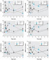
Figure 3
Figure 3
Time-dependent changes in pERK elicited by combinations of BPA at two different environmentally relevant concentrations. GH3/B6/F10 cells were cotreated with 10−14 M BPA (AC) or 10−9 M BPA (DF) plus 1 nM E2 (A,D), E1 (B,E), or E3 (C,F). The pERK levels were measured after different times of cotreatment. *p < 0.05 compared with vehicle-treated control cells, shaded to match each set of data symbols (*, formula image, ✫). #p < 0.05 compared with cells treated with 1 nM E2, E1, or E3 alone.
Figure 4
Figure 4
Concentration-dependent changes in pERK caused by short-chain alkylphenols. Cells were treated for 5 min with a combination of 1 nM E2 (A,D), E1 (B,E), or E3 (C,F) plus different concentrations of EP (AC) or PP (DF), and pERK was assayed. The blue horizontal bar indicates the pERK level and error range in vehicle-treated cells (V); the crosshatched horizontal bar indicates the pERK value in cells treated with nanomolar concentrations of E2, E1, or E3 alone. *p < 0.05 compared with vehicle-treated cells. #p < 0.05 compared with cells treated with E2, E1, or E3 alone.
Figure 5
Figure 5
Concentration-dependent changes in pERK caused by the long-chain alkylphenols. Cells were treated for 5 min with a combination of 1 nM E2 (A,D), E1 (B,E), or E3 (C,F) plus different concentrations of OP (AD) or NP (DF), and pERK was assayed. The blue horizontal bar indicates the pERK level and error range in vehicle-treated cells (V); the crosshatched horizontal bar indicates the pERK value in cells treated with nanomolar concentrations of E2, E1, or E3 alone. *p < 0.05 compared with vehicle-treated cells. #p < 0.05 compared with cells treated with E2, E1, or E3 alone.
Figure 6
Figure 6
Concentration-dependent alteration of physiologic estrogen-induced pERK by BPA. Cells were treated for 5 min with different concentrations of BPA with or without 1 nM E2 (A), E1 (B), or E3 (C). The blue horizontal bar indicates the pERK level and error range in vehicle-treated cells (V); the crosshatched horizontal bar indicates the pERK value in cells treated with nanomolar concentrations of E2, E1, or E3 alone. *p < 0.05 compared with vehicle-treated cells. #p < 0.05 compared with cells treated with E2, E1, or E3 alone.
Figure 7
Figure 7
Concentration dependence of pERK (A–D) and proliferation (E–H) on selective ER agonists. pERK was measured in cells after 5 min of treatment with different concentrations of E2 (A), the ERα agonist PPT (B), the ERβ agonist DPN (C), or the GPER agonist G1 (D). Proliferation was measured (via the crystal violet assay) in cells treated for 3 days with matching concentrations of E2 (E), PPT (F), DPN (G), or G1 (H). V, vehicle-treated cells. *p < 0.05 compared with vehicle-treated cells.

Comment in

Similar articles

Cited by

  • Suppression of lymphoma growth by the xenoestrogens bisphenol A and genistein.
    Yakimchuk K, Bangalore Revanna C, Huang D, Inzunza J, Okret S. Yakimchuk K, et al. Endocr Connect. 2018 Dec;7(12):1472-1479. doi: 10.1530/EC-18-0459. Endocr Connect. 2018. PMID: 30496125 Free PMC article.
  • Assessing the carcinogenic potential of low-dose exposures to chemical mixtures in the environment: the challenge ahead.
    Goodson WH 3rd, Lowe L, Carpenter DO, Gilbertson M, Manaf Ali A, Lopez de Cerain Salsamendi A, Lasfar A, Carnero A, Azqueta A, Amedei A, Charles AK, Collins AR, Ward A, Salzberg AC, Colacci A, Olsen AK, Berg A, Barclay BJ, Zhou BP, Blanco-Aparicio C, Baglole CJ, Dong C, Mondello C, Hsu CW, Naus CC, Yedjou C, Curran CS, Laird DW, Koch DC, Carlin DJ, Felsher DW, Roy D, Brown DG, Ratovitski E, Ryan EP, Corsini E, Rojas E, Moon EY, Laconi E, Marongiu F, Al-Mulla F, Chiaradonna F, Darroudi F, Martin FL, Van Schooten FJ, Goldberg GS, Wagemaker G, Nangami GN, Calaf GM, Williams G, Wolf GT, Koppen G, Brunborg G, Lyerly HK, Krishnan H, Ab Hamid H, Yasaei H, Sone H, Kondoh H, Salem HK, Hsu HY, Park HH, Koturbash I, Miousse IR, Scovassi AI, Klaunig JE, Vondráček J, Raju J, Roman J, Wise JP Sr, Whitfield JR, Woodrick J, Christopher JA, Ochieng J, Martinez-Leal JF, Weisz J, Kravchenko J, Sun J, Prudhomme KR, Narayanan KB, Cohen-Solal KA, Moorwood K, Gonzalez L, Soucek L, Jian L, D'Abronzo LS, Lin LT, Li L, Gulliver L, McCawley LJ, Memeo L, Vermeulen L, Leyns L, Zhang L, Valverde M, Khatami M, Romano MF, Chapellier M, Williams MA, Wade M, Manjili MH, Lleonart ME, Xia M, Gonzalez MJ, Karamouzis … See abstract for full author list ➔ Goodson WH 3rd, et al. Carcinogenesis. 2015 Jun;36 Suppl 1(Suppl 1):S254-96. doi: 10.1093/carcin/bgv039. Carcinogenesis. 2015. PMID: 26106142 Free PMC article. Review.
  • Neurological Effects of Bisphenol A and its Analogues.
    Inadera H. Inadera H. Int J Med Sci. 2015 Oct 30;12(12):926-36. doi: 10.7150/ijms.13267. eCollection 2015. Int J Med Sci. 2015. PMID: 26664253 Free PMC article. Review.
  • Endocrine-disrupting chemicals: associated disorders and mechanisms of action.
    De Coster S, van Larebeke N. De Coster S, et al. J Environ Public Health. 2012;2012:713696. doi: 10.1155/2012/713696. Epub 2012 Sep 6. J Environ Public Health. 2012. PMID: 22991565 Free PMC article. Review.
  • Endocrine disruption via estrogen receptors that participate in nongenomic signaling pathways.
    Watson CS, Jeng YJ, Guptarak J. Watson CS, et al. J Steroid Biochem Mol Biol. 2011 Oct;127(1-2):44-50. doi: 10.1016/j.jsbmb.2011.01.015. Epub 2011 Feb 12. J Steroid Biochem Mol Biol. 2011. PMID: 21300151 Free PMC article. Review.

References

    1. Alonso-Magdalena P, Morimoto S, Ripoll C, Fuentes E, Nadal A. The estrogenic effect of bisphenol A disrupts pancreatic β-cell function in vivo and induces insulin resistance. Environ Health Perspect. 2006;114:106–112. - PMC - PubMed
    1. Alyea RA, Laurence SE, Kim SH, Katzenellenbogen BS, Katzenellenbogen JA, Watson CS. The roles of membrane estrogen receptor subtypes in modulating dopamine transporters in PC-12 cells. J Neurochem. 2008;106:1525–1533. - PMC - PubMed
    1. Alyea RA, Watson CS. Differential regulation of dopamine transporter function and location by low concentrations of environmental estrogens and 17β-estradiol. Environ Health Perspect. 2009a;117:778–783. - PMC - PubMed
    1. Alyea RA, Watson CS. Nongenomic mechanisms of physiological estrogen-mediated dopamine efflux. BMC Neurosci. 2009b;10:59. doi: 10.1186/1471-2202-10-59. [Online 16 June 2009[ - DOI - PMC - PubMed
    1. Belcheva MM, Coscia CJ. Diversity of G protein- coupled receptor signaling pathways to ERK/MAP kinase. Neurosignals. 2002;11:34–44. - PMC - PubMed

Publication types

MeSH terms

LinkOut - more resources