. 2010 Nov 26;285(48):37641-9.
doi: 10.1074/jbc.M110.132415. Epub 2010 Sep 24.

Identification of a lysosomal pathway regulating degradation of the bone morphogenetic protein receptor type II

Affiliations

Identification of a lysosomal pathway regulating degradation of the bone morphogenetic protein receptor type II

Hannah J Durrington et al. J Biol Chem. .

Abstract

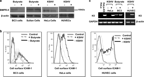
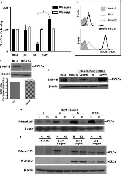
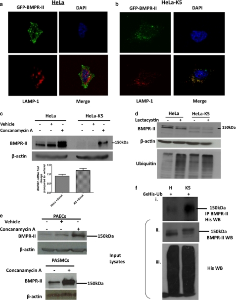
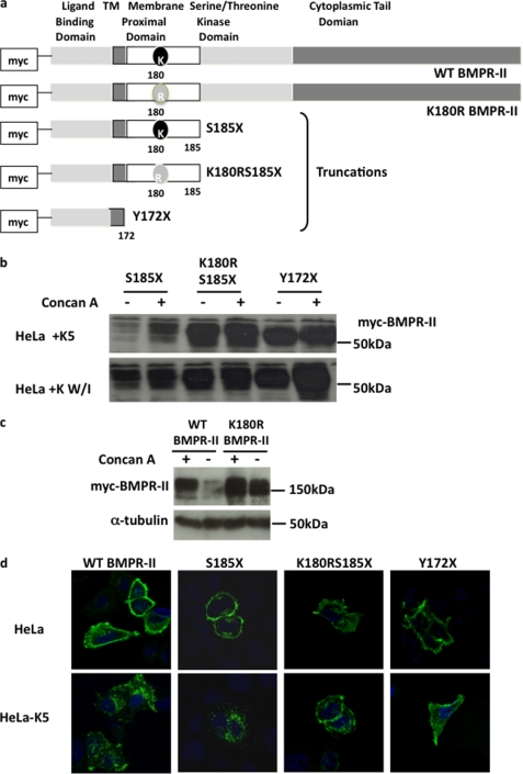
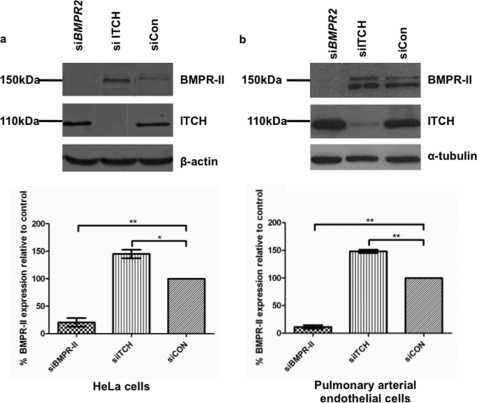
Bone morphogenetic proteins (BMPs) are critically involved in early development and cell differentiation. In humans, dysfunction of the bone morphogenetic protein type II receptor (BMPR-II) is associated with pulmonary arterial hypertension (PAH) and neoplasia. The ability of Kaposi sarcoma-associated herpesvirus (KSHV), the etiologic agent of Kaposi sarcoma and primary effusion lymphoma, to down-regulate cell surface receptor expression is well documented. Here we show that KSHV infection reduces cell surface BMPR-II. We propose that this occurs through the expression of the viral lytic gene, K5, a ubiquitin E3 ligase. Ectopic expression of K5 leads to BMPR-II ubiquitination and lysosomal degradation with a consequent decrease in BMP signaling. The down-regulation by K5 is dependent on both its RING domain and a membrane-proximal lysine in the cytoplasmic domain of BMPR-II. We demonstrate that expression of BMPR-II protein is constitutively regulated by lysosomal degradation in vascular cells and provide preliminary evidence for the involvement of the mammalian E3 ligase, Itch, in the constitutive degradation of BMPR-II. Disruption of BMP signaling may therefore play a role in the pathobiology of diseases caused by KSHV infection, as well as KSHV-associated tumorigenesis and vascular disease.


Figures

FIGURE 1.
FIGURE 1.
KSHV infection causes a reduction in BMPR-II protein. a, immunoblot for BMPR-II protein in BC3 cells and control Sultan cells before and after treatment with 2 mm sodium butyrate for 24 h. BMPR-II levels in HeLa cells 2 days after KSHV infection and also in HUVECS 7 days after KSHV infection when compared with uninfected control cells are shown. b, flow cytometry for cell surface ICAM-1, a known target of K5, in BC3 cells after induction of lytic KSHV and in HeLa cells infected with KSHV after 2 days and in HUVECs 7 days after infection. Controls in these experiments were uninduced BC3 cells and uninfected HeLa and HUVEC cells stained with secondary antibody only. c, RT-PCR analysis for K5 mRNA from KSHV-infected HeLa cells when compared with HeLa controls and BC3 cells before and after induction of lytic KSHV infection with butyrate and in control and KSHV-infected HUVECs. Control HeLa cells and stably transfected HeLa-K5 cells served as negative and positive controls for the PCR, respectively. Representative images of three experiments are shown.
FIGURE 2.
FIGURE 2.
K5 specifically reduces BMPR-II, a process that is dependent on the RING domain of K5. a, cell surface binding of radiolabeled BMP4 in HeLa cells stably expressing K5 (HeLa-K5) when compared with control HeLa cells and HeLa cells stably expressing K3 (a homolog of K5) (HeLa-K3) and in HeLa cells expressing a mutated, non-functioning K5 (K5W). Radiolabeled TGF-β1 binding is also shown. (*, p < 0.01). Error bars indicate S.E. b, flow cytometry for cell surface BMPR-II and ICAM-1 expression in HeLa and HeLa-K5 cells. Controls were incubated with secondary antibody alone. Error bars indicate S.E. c, immunoblot for BMPR-II protein in HeLa and HeLa-K5 cells showing reduced BMPR-II protein in HeLa-K5 cells. Graph shows similar levels of BMPR-II mRNA transcripts in both cell lines. d, immunoblot for BMPR-II protein levels in control HeLa cells, stably expressing HeLa-K5 cells and HeLa cells transiently transfected with K5 or a non-functioning RING mutant of K5 (K5W/I). e, immunoblot showing the level of Smad 1 phosphorylation (P-Smad 1/5) following BMP4 stimulation in HeLa and HeLa-K5 cells. f, immunoblot of the TGF-β downstream signaling molecule, phospho-Smad 2 (P-Smad 2), in HeLa and HeLa-K5 cells following TGF-β1 stimulation. Studies shown are representative of 3 independent experiments.
FIGURE 3.
FIGURE 3.
K5 causes redistribution of BMPR-II from the cell surface to a lysosomal compartment. a and b, confocal microscopy showing the localization of GFP-tagged BMPR-II (green), the lysosomal marker, LAMP-1 (red), nuclear DAPI stain (blue), and merged images in HeLa (a) and HeLa-K5 (b) cells. c, immunoblot showing BMPR-II protein levels before and after treatment of HeLa and HeLa-K5 cells with concanamycin A (lysosomal inhibitor) (vehicle is ethanol) and graph of BMPR-II mRNA transcript levels under the same conditions. Error bars indicate S.E. d, immunoblots for BMPR-II and endogenous ubiquitin levels in the presence and absence of lactacystin (proteasomal inhibitor). e, immunoblots for BMPR-II protein in pulmonary arterial endothelial cells (PAECs) and pulmonary arterial smooth muscle cells (PASMCs) before and after treatment with concanamycin A. f, panel i, viral transduction of HeLa and HeLa-K5 cells with a His6-tagged ubiquitin (6xHis-Ub) followed by a BMPR-II immunoprecipitation (IP) and His Western blot. f, panels ii and iii, Western blots (WB) show the presence of endogenous BMPR-II in input lysates (panel ii) and also the presence of transduced His6 ubiquitin (panel iii). Studies shown are representative of 3 independent experiments.
FIGURE 4.
FIGURE 4.
K5 targets the membrane-proximal lysine of BMPR-II (Lys-180). a, a series of N-terminally Myc-tagged BMPR-II constructs were made: full-length wild type BMPR-II, full-length BMPR-II in which the lysine at position 180 was mutated to arginine, truncated (S185X) BMPR-II, truncated BMPR-II in which the lysine at 180 was mutated to arginine (K180R/S185X), and BMPR-II in which the entire intracellular domain was missing (Y172X). TM, transmembrane. b, immunoblot for Myc protein in HeLa cells following transient transfection with the Myc-tagged BMPR-II tail truncation constructs and either K5 or the K5 RING mutant, K5W/I, before and after concanamycin A (Concan A) treatment. c, immunoblot for Myc protein in HeLa and HeLa-K5 cells after transfection with the full-length constructs, Myc-BMPR-II or Myc-K180R, with and without pretreatment with concanamycin A. d, confocal images were generated after staining for Myc in HeLa and HeLa-K5 cells after transient transfection with the Myc-tagged BMPR-II tail truncated constructs (magnification ×60 oil immersion objective). In d, all blots and images are representative of 3 independent experiments.
FIGURE 5.
FIGURE 5.
siRNA knockdown of Itch causes an increase in endogenous levels of BMPR-II. a, immunoblots for BMPR-II protein and Itch, demonstrating the effect of BMPR-II or ITCH knockdown in HeLa cells, when compared with control siRNA (siCON). b, similar experiments in pulmonary arterial endothelial cells. Graphs show results of densitometry from n = 3–4 independent experiments (*, p < 0.05 and **, p < 0.01). Error bars indicate S.E.

References

    1. Miyazono K., Maeda S., Imamura T. (2005) Cytokine Growth Factor Rev. 16, 251–263 - PubMed
    1. Beppu H., Kawabata M., Hamamoto T., Chytil A., Minowa O., Noda T., Miyazono K. (2000) Dev. Biol. 221, 249–258 - PubMed
    1. Liu D., Wang J., Kinzel B., Müeller M., Mao X., Valdez R., Liu Y., Li E. (2007) Blood 110, 1502–1510 - PubMed
    1. International, PPH Consortium Lane K. B., Machado R. D., Pauciulo M. W., Thomson J. R., Philips J. A., 3rd, Loyd J. E., Nichols W. C., Trembath R. C. (2000) Nat. Genet. 26, 81–84 - PubMed
    1. Thomson J. R., Machado R. D., Pauciulo M. W., Morgan N. V., Humbert M., Elliott G. C., Ward K., Yacoub M., Mikhail G., Rogers P., Newman J., Wheeler L., Higenbottam T., Gibbs J. S., Egan J., Crozier A., Peacock A., Allcock R., Corris P., Loyd J. E., Trembath R. C., Nichols W. C. (2000) J. Med. Genet. 37, 741–745 - PMC - PubMed

Publication types

MeSH terms