. 2010 Nov 26;285(48):37725-32.
doi: 10.1074/jbc.M110.139527. Epub 2010 Sep 24.

The MBT repeats of L3MBTL1 link SET8-mediated p53 methylation at lysine 382 to target gene repression

Affiliations

The MBT repeats of L3MBTL1 link SET8-mediated p53 methylation at lysine 382 to target gene repression

Lisandra E West et al. J Biol Chem. .

Abstract

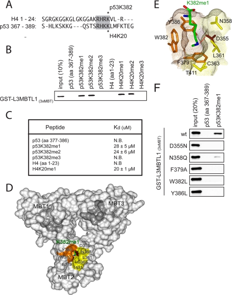
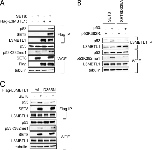
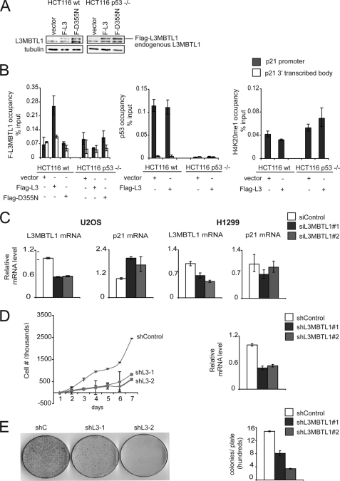
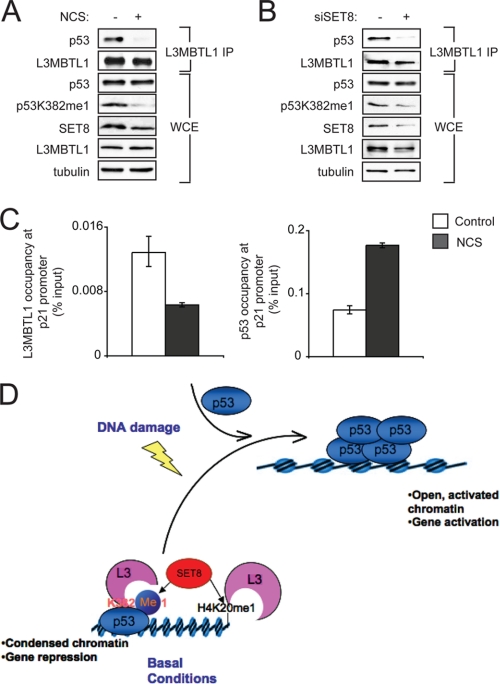
The p53 tumor suppressor protein is regulated by multiple post-translational modifications, including lysine methylation. We previously found that monomethylation of p53 at lysine 382 (p53K382me1) by the protein lysine methyltransferase (PKMT) SET8/PR-Set7 represses p53 transactivation of target genes. However, the molecular mechanism linking p53K382 monomethylation to repression is not known. Here we show in biochemical and crystallographic studies the preferential recognition of p53K382me1 by the triple malignant brain tumor (MBT) repeats of the chromatin compaction factor L3MBTL1. We demonstrate that SET8-mediated methylation of p53 at Lys-382 promotes the interaction between L3MBTL1 and p53 in cells, and the chromatin occupancy of L3MBTL1 at p53 target promoters. In the absence of DNA damage, L3MBTL1 interacts with p53K382me1 and p53-target genes are repressed, whereas depletion of L3MBTL1 results in a p53-dependent increase in p21 and PUMA transcript levels. Activation of p53 by DNA damage is coupled to a decrease in p53K382me1 levels, abrogation of the L3MBTL1-p53 interaction, and disassociation of L3MBTL1 from p53-target promoters. Together, we identify L3MBTL1 as the second known methyl-p53 effector protein, and provide a molecular explanation for the mechanism by which p53K382me1 is transduced to regulate p53 activity.


Figures

FIGURE 1.
FIGURE 1.
L3MBTL1(3xMBT) recognizes p53 monomethylated at lysine 382 (p53K382me1) in vitro. A, sequence alignment of the H4 N terminus and p53 C-terminal tail. Homology surrounding the SET8/53BP1 binding site is highlighted. Asterisks indicate methylation sites. B, L3MBTL1(3xMBT) binds to p53K382me1 and p53K382me2 peptides. Shown are the results from Western analysis of peptide pull-down assays with the indicated biotinylated peptides and GST-L3MBTL1(3xMBT). C, dissociation constants (Kd) of p53 or histone H4 peptides for L3MBTL1(3xMBT) were determined by ITC. Kd of H4K20me1 is shown as control. D, L3MBTL1(3xMBT) repeats are shown as a solid surface with the residues comprising the p53K382me1-binding pocket of MBT2 labeled and colored orange, brown, and yellow for aromatic, acidic, and hydrophobic/polar residues, respectively. Lysine 382me1 of the p53K382me1 peptide is shown as a stick model with C, O, and N atoms colored green, red, and blue, respectively. The unoccupied semi-aromatic pockets MBT1 and MBT3 are colored gray. E, close-up view of the K382me1 binding cage. A dotted red line depicts the hydrogen bond between the methylammonium proton and the carboxyl group of Asp-355. F, point mutations in the MBT2 repeat of L3MBTL1 abolish the p53K382me1 binding activity of L3MBTL1(3xMBT) in vitro. Western analysis of the indicated GST-L3MBTL1(3xMBT) mutants in peptide pull-down assays are shown.
FIGURE 2.
FIGURE 2.
L3MBTL1 binds to p53K382me1 in vivo. A, SET8 augments the interaction between p53 and L3MBTL1 in vivo. Western analysis with the indicated antibodies of the Flag IPs or whole cell extract (WCE) from 293T cells expressing the indicated proteins is shown. Tubulin levels in the WCE are shown as loading control. B, monomethylation on p53K382 by SET8 is important for the association of L3MBTL1 and p53 in vivo. Western analysis with the indicated antibodies of endogenous L3MBTL1 IPs or WCE from H1299 cells expressing wild-type p53 or p53K382R mutant in the presence of SET8 or SET8(D338A) as indicated. Tubulin is shown as a loading control. C, intact MBT2 domain is required for interaction between L3MBTL1 and p53 in vivo. Western analysis with the indicated antibodies of the Flag IPs or WCE from 293T cells expressing wild-type F-L3MBTL1 or F-L3MBTL1(D355N) mutant ± SET8. Tubulin levels in the WCE are shown as loading control.
FIGURE 3.
FIGURE 3.
L3MBTL1 represses p53 target genes under normal conditions. A, Western blot with L3MBTL1 antibodies of WCE from the indicated cell lines transduced with control, Flag-L3MBTL1, or Flag-L3MBTL1-D355N retrovirus. B, p53 stabilizes L3MBTL1 at target genes under basal conditions. Chromatin immunoprecipitation (ChIP) assays to determine occupancy at the promoter and 3′ transcribed body region of the p21 gene in wt and p53−/− HCT116 cells stably expressing Flag-L3MBTL1 or Flag-L3MBTL1-(D355N) as indicated. ChIPs: Flag (left), p53 (middle), H4K20me1 (right). Occupancy values (% input) were determined by real-time PCR. C, p21 expression increases upon L3MBTL1 depletion in the presence of endogenous p53 and in the absence of DNA damage. U2OS or the p53-negative cell line H1299 treated with control or two independent L3MBTL1 siRNAs and L3MBTL1 and p21 mRNA levels were determined by real-time PCR. D, L3MBTL1 depletion attenuates cellular proliferation rates. Left panel, 7-day growth curve time course for U2OS cells stably expressing control or two independent L3MBTL1 shRNAs. Error bars indicate S.E. from three experiments. Right panel, real-time PCR of L3MBTL1 mRNA levels of cells used in the growth curve experiment. E, L3MBTL1 depletion attenuates colony growth. Left panel, colony formation panel of cells as in D. Representative plates are shown for control and the two L3MBTL1 stable knockdown lines. Right panel, quantitation of colonies counted for each cell line. Error bars indicate S.E. from three independent experiments.
FIGURE 4.
FIGURE 4.
The endogenous interaction between L3MBTL1 and p53 is disrupted upon DNA damage. A, Western analysis with the indicated antibodies of L3MBTL1 IPs or WCE prepared from 293T cells, ± 0.5 μg/ml the radiomimetic NCS. Note that p53 levels do not increase upon DNA damage in 293T cells. B, SET8 depletion abrogates the interaction between endogenous L3MBTL1 and p53. Western analysis with the indicated antibodies of the L3MBTL1 IPs or WCE prepared from 293T cells ± SET8 siRNA (11). C, L3MBTL1 occupancy at the p21 promoter decreases upon DNA damage. ChIP assays of endogenous L3MBTL1 at the p21 promoter in U2OS cells ± treatment with 0.5 μg/ml NCS. Occupancy values (% input) were determined by real-time PCR. D, model for SET8-mediated repression of p53 through promotion of p53-L3MBTL1 binding. In the absence of DNA damage, SET8 generates a population of p53 monomethylated at Lys-382. p53K382me1 binds to and stabilizes chromatin compaction factor L3MBTL1 at the promoter regions of p53 target genes, which results in a repressive chromatin state. Upon DNA damage, Lys-382 monomethylation levels decrease by as yet unknown mechanism (possibly via demethylation, higher methylation of p53K382, or simply is diluted out by the newly stabilized p53 protein) and this leads to L3MBTL1 dissociation from p53-target genes and a chromatin environment favorable for active gene transcription.

References

    1. Shi Y., Whetstine J. R. (2007) Mol. Cell. 25, 1–14 - PubMed
    1. Sims R. J., 3rd, Reinberg D. (2006) Genes Dev. 20, 2779–2786 - PubMed
    1. Bannister A. J., Kouzarides T. (2004) Methods Enzymol. 376, 269–288 - PubMed
    1. Daniel J. A., Pray-Grant M. G., Grant P. A. (2005) Cell Cycle. 4, 919–926 - PubMed
    1. Baker L. A., Allis C. D., Wang G. G. (2008) Mutat. Res. 647, 3–12 - PMC - PubMed

Publication types

MeSH terms

Associated data