. 2011 Feb 15;183(4):511-21.
doi: 10.1164/rccm.201005-0744OC. Epub 2010 Sep 24.

Conditional depletion of airway progenitor cells induces peribronchiolar fibrosis

Affiliations

Conditional depletion of airway progenitor cells induces peribronchiolar fibrosis

Anne-Karina T Perl et al. Am J Respir Crit Care Med. .

Abstract

Rationale: The respiratory epithelium has a remarkable capacity to respond to acute injury. In contrast, repeated epithelial injury is often associated with abnormal repair, inflammation, and fibrosis. There is increasing evidence that nonciliated epithelial cells play important roles in the repair of the bronchiolar epithelium after acute injury. Cellular processes underlying the repair and remodeling of the lung after chronic epithelial injury are poorly understood.

Objectives: To identify cell processes mediating epithelial regeneration and remodeling after acute and chronic Clara cell depletion.

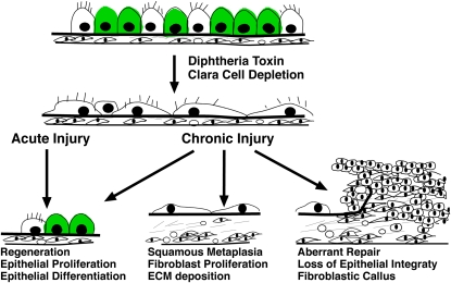
Methods: A transgenic mouse model was generated to conditionally express diphtheria toxin A to ablate Clara cells in the adult lung. Epithelial regeneration and peribronchiolar fibrosis were assessed after acute and chronic Clara cell depletion.

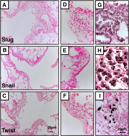
Measurements and main results: Acute Clara cell ablation caused squamous metaplasia of ciliated cells and induced proliferation of residual progenitor cells. Ciliated cells in the bronchioles and pro-surfactant protein C-expressing cells in the bronchiolar alveolar duct junctions did not proliferate. Epithelial cell proliferation occurred at multiple sites along the airways and was not selectively associated with regions around neuroepithelial bodies. Chronic Clara cell depletion resulted in ineffective repair and caused peribronchiolar fibrosis.

Conclusions: Colocalization of proliferation and cell type-specific markers demonstrate that Clara cells are critical airway progenitor cells. Continuous depletion of Clara cells resulted in persistent squamous metaplasia, lack of normal reepithelialization, and peribronchiolar fibrosis. Induction of proliferation in subepithelial fibroblasts supports the concept that chronic epithelial depletion caused peribronchiolar fibrosis.


Figures

Figure 1.
Figure 1.
Clara cell depletion after acute doxycycline (dox) exposure: Hematoxylin and eosin staining of histological lung sections after (A and D) 2 days of dox exposure and (B and E) 2 days of dox exposure followed by 2 days of recovery in triple transgenic mice from (AC) Scgb1a1rtTA line 1 and (DF) Scgb1a1rtTA line 2. Immunohistochemistry for the proliferation marker phosphohistone H3 (pH3) revealed increased proliferation in Scgb1a1rtTA line 1 (inset in B); (C and F) immunohistochemistry using goat anti–Clara cell secretory protein (CCSP) revealed that 95.5 ± 2.0% of Clara cells were depleted after 2 days of dox exposure and 2 days of recovery. Arrows in B and C indicate bronchiolar hyperproliferation; arrows in E and F indicate clusters of cuboidal cells; asterisks in E show cells sloughing off; the arrowhead in F shows a bronchiole depleted of Clara cells. Figures are representative of at least five individual mice per genotype and at each time. Scale bars: (A, B, D, and E) 20 μm; (C and F) 500 μm.
Figure 2.
Figure 2.
Residual squamous cells maintain the bronchiolar epithelium. (A, E, and I) Triple transgenic mice were exposed to doxycycline (dox) for 2 days to activate diphtheria toxin A (DT-A) by Cre recombination. Histology was assessed (B, F, and J) 2 days, (C, G, and K) 5 days, and (D, H, and L) 10 days after dox withdrawal, using (AD) hematoxylin and eosin (H&E) staining. (EH) Rabbit anti–Clara cell secretory protein (CCSP), (F) a more sensitive goat anti-CCSP (inset), and (IL) β-tubulin antibodies were used to stain the tissue. After 2 days of dox, residual cuboidal cells in the bronchioles express high levels of CCSP (E, arrowhead). Two days after dox withdrawal only 4.5 ± 2% of cells express CCSP; expression in these cells is low and can be detected only with the more sensitive goat anti-CCSP antibody. Between 5 and 10 days after dox withdrawal, the percentage of CCSP-positive cells increased to 45.5 ± 16.2% and the level of CCSP expression per cell increased (G and H, arrowheads). In the absence of cuboidal cells, squamous ciliated cells (arrowheads in IL) covered the bronchioles. Results are representative of n ≥ 3 triple transgenic Scgb1a1/DT-A mice.
Figure 3.
Figure 3.
Cell proliferation during repair of the bronchiolar epithelium. Mice were treated with doxycycline (dox) for 2 days; proliferation was assessed after 2 days of dox treatment and 2 and 5 days of recovery, using immunohistochemistry for the proliferation marker Ki67. After 2 days of recovery 8.5 ± 4.4% of the cells were proliferative and 10.0 ± 7.7% were proliferative after 5 days. Immunofluorescence images were merged with images acquired using differential interference contrast optics: goat anti–Clara cell secretory protein (CCSP) (green in A, D, and G; red in C, F, and I), Ki67 (pink), forkhead-box J1 (FOXJ1; green). Ki67 expression was colocalized with CCSP in Clara cells but not with FOXJ1 staining (ciliated cells). Colocalization of CCSP and FOXJ1 showed a subset of dual-positive cells at 5 days but not at 2 days of recovery. Arrows, double-positive cells; asterisk, rare proliferating cuboidal cell that lacks CCSP. Results are representative of n ≥ 3 triple transgenic Scgb1a1/DT-A mice.
Figure 4.
Figure 4.
Assessment of proliferation in neuroepithelial body (NEB) and bronchiolar alveolar duct junction (BADJ) regions. Immunohistochemistry was performed for Clara cell secretory protein (CCSP) (green in A, red in EH), calcitonin gene–related peptide (CGRP) (red in A, green in B), Ki67 (red in B), and pro–surfactant protein C (SPC) (green in EH). (A and B) NEB regions were determined by positive CGRP staining and included bronchiolar cells covering the NEB and two or three cells outside the CGRP-positive area. After 2 days of doxycycline (dox) treatment and 5 days of recovery, not all NEB regions were covered with regenerating Clara cells; proliferating cells were found outside of NEB regions. (C and D) Sections containing all five lobes were double labeled for Ki67 and CGRP, and counterstained with 4′,6-diamidino-2-phenylindole (DAPI). The number of DAPI-positive cells per millimeter of airway and the percentage of KI67-positive cells per DAPI-positive cell in NEB and non-NEB regions were determined. (C) Cell density around NEB regions (solid columns) was 112 cells/mm of airway after 2 days of recovery and 143 cells/mm after 5 days of recovery, which was significantly more than the 66 and 81 cells/mm in the non-NEB regions (open columns) (P < 0.018 and P < 0.045, respectively). (D) After 2 days of recovery proliferation in NEB (solid columns; 9.5%) and non-NEB (open columns; 8.5%) regions was comparable (P < 0.155). After 5 days of recovery proliferation in NEB (black; 14.7%) was slightly increased when compared with non-NEB regions (white; 10.0%) (P < 0.029). Results are expressed as the means ± SE of six sections of three to five animals per group. (EH) Colocalization of goat anti-CCSP (red) and SPC (green) in the bronchiolar epithelium was assessed after acute injury and recovery for 2, 5, and 10 days. Immunofluorescence images were merged with images acquired with differential interference contrast optics. Only after 2 days of recovery were rare clusters of dual-positive cells found in BADJ regions (1 cluster per 15 BADJ regions). High magnification of a dual-positive BADJ cluster is shown in the inset in (F). Results are representative of more than 45 BADJ regions from 3 sections, containing all 5 lobes, of 3–5 triple transgenic Scgb1a1/DT-A animals per group at each time point.
Figure 5.
Figure 5.
Chronic loss and aberrant repair of Clara cells after continuous diphtheria toxin A (DT-A) expression. Immunohistochemistry for Clara cell secretory protein (CCSP) was assessed on lung sections of triple transgenic Scgb1a1/DT-A mice after 2 days (AC) or 10 days of DT-A expression (DI), and recovery for (B and E) 10 days, (C and F) 3 weeks, and (GI) 19 weeks. After 3 weeks of recovery Clara cells lined many bronchioles (arrows) but were absent in some bronchiolar regions. After 19 weeks of recovery, some bronchioles still lacked Clara cells (arrowheads in BG). Peribronchiolar fibrosis was found after 10 days of doxycycline (dox) treatment (insets in D and E) and persisted throughout 19 weeks of recovery (arrows in H). After 19 weeks of recovery, aberrant epithelial repair was evident (H). Scale bars: (AF) 200 μm; (G and H) 100 μm; (I) 5 μm. Results are representative of n ≥ 3 triple transgenic Scgb1a1/DT-A mice.
Figure 6.
Figure 6.
Extent of Clara cell depletion and mesenchymal fibrosis. Random sections from two or three animals and containing all five lobes were stained for Clara cell secretory protein (CCSP). Pictures of all airways were taken. (A) The length of total airways and CCSP-positive airways was determined. Clara cell depletion was expressed as the percentage of airways covered with CCSP-positive cells. In airways with no injury CCSP stain covered 100% of the airways. After 2 days of doxycycline (dox) and 2 days off dox treatment only 4.5 ± 2.0% of the airways were covered with Clara cells. After 10 days Clara cells significantly regenerated to cover 45.5 ± 16.3% of the bronchioles (P < 0.001) and continued to significantly regenerate to cover 70.6 ± 4.7% after 21 days of recovery (P < 0.001). After 10 days of dox 16.5 ± 8.6% of the airways were CCSP positive, which is comparable to denudation after 2 days (P < 0.06). Ten days after chronic injury Clara cells regenerated and covered 65.7 ± 11.6% (P < 0.001) of the airways. Thereafter the percentage of Clara cells did not increase (57.7 ± 6.9%; P < 0.20). (B) The area of mesenchymal tissue adjacent to the bronchiolar basement membrane was determined in uninjured bronchioles and after 10 days of dox exposure. The extent of fibrosis was determined as millimeters squared per millimeter of airway. In uninjured animals the average size of the underlying mesenchyme was 4.8 ± 0.3 mm2/mm of airway. After 10 days of injury the mesenchyme increased in thickness to 12.6 ± 2.0 mm2/mm (P < 0.001) and remained at 14.1 ± 0.2 and 14.4 ± 0.1 mm2/mm after 10 and 21 days of repair (P < 0.39). Results are expressed as means ± SE of 80–160 mm of airway from two or three animals per group.
Figure 7.
Figure 7.
Chronic Clara cell depletion results in peribronchiolar fibrosis. After 10 days of doxycycline (dox) treatment, immunohistochemistry on lung sections of triple transgenic mice for cell type–specific markers was performed with (A) goat anti–Clara cell secretory protein (CCSP), (B) β-tubulin, (D) platelet–endothelial cell adhesion molecule (PECAM), and (F) α-smooth muscle actin (α-SMA). Proliferation was assessed by (C) Ki67 staining and extracellular matrix deposition was assessed by (E) trichrome staining. Peribronchiolar fibrosis was associated with loss of Clara cells, proliferating fibroblasts, and deposition of extracellular matrix but was not associated with smooth muscle hyperplasia. Results are representative of n ≥ 3 triple transgenic Scgb1a1/DT-A mice.
Figure 8.
Figure 8.
Immunohistochemistry (IHC) for epithelial and stromal cell markers in regions of peribronchiolar fibrosis. IHC was performed for (A) pan-cytokeratin, (B) N-cadherin, (C) collagen IV, and (D) desmin, and dual immunofluorescence was performed for thyroid transcription factor (TTF)-1 and the smooth muscle cell marker α-smooth muscle actin (α-SMA) (E) or the mesenchymal marker vimentin (F). A squamous layer of pan-cytokeratin–positive epithelial cells lined the bronchioles. Weak and patchy expression of TTF-1 suggested loss of epithelial differentiation; lack of desmin and α-SMA expression demonstrated that fibrosis was not due to myofibroblast hypertrophy. Arrows, TTF-1 expression in alveolar type II cells; arrowheads, low TTF-1 expression in squamous cells. Results are representative of n ≥ 3 triple transgenic Scgb1a1/DT-A mice.
Figure 9.
Figure 9.
Immunohistochemistry for epithelial and stromal markers in regions of fibromyxoid plugs. Immunohistochemistry was performed for (A) pan-cytokeratin, (B) N-cadherin, (C) collagen IV, and (D) desmin, and dual immunofluorescence was performed for thyroid transcription factor (TTF)-1 and the smooth muscle cell marker α-smooth muscle actin (α-SMA) (E) or the mesenchymal marker vimentin (F). The epithelial layer was interrupted in regions of fibrotic callus formation, and fibrotic lesions contained vimentin-positive, α-SMA–negative fibroblasts. Arrows, TTF-1 expression in alveolar type II cells; arrowhead, low TTF-1 expression in squamous cells. Results are representative of n ≥ 3 triple transgenic Scgb1a1/DT-A mice.
Figure 10.
Figure 10.
Lack of epithelial–mesenchymal transition (EMT) markers in fibrotic lesions. Immunohistochemistry for (A, D, and G) SLUG, (B, E, and H) SNAIL, and (C, F, and I) TWIST was performed on lung sections of triple transgenic mice after 10 days of diphtheria toxin A (DT-A) expression (AF) and on urethane-induced lung tumors (GI), which were used as positive controls for the antibody staining. SLUG, SNAIL, and TWIST staining was not detected in areas with peribronchiolar fibrosis or in fibrotic calluses. Results are representative of n ≥ 3 triple transgenic Scgb1a1/DT-A mice at all acute and chronic injury and recovery time points.
Figure 11.
Figure 11.
Schematic process of lung injury and repair after acute and chronic Clara cell depletion. Diphtheria toxin A (DT-A) was expressed in Clara cells after doxycycline (dox) treatment causing death of nonciliated cuboidal cells. After acute epithelial injury (2 d of DT-A expression), the remaining ciliated cells were squamous and covered the basement membrane. From 2 to 10 days after cessation of DT-A expression, the bronchiolar epithelium proliferated and differentiated. Acute injury did not cause stromal thickening. In contrast, after chronic injury (10 d of DT-A expression) squamous metaplasia and peribronchiolar fibrosis were observed. Squamous metaplasia, fibroblast proliferation, and excessive matrix deposition were associated with peribronchiolar fibrosis. Regional loss of epithelial integrity was associated with intrabronchiolar lesions, ineffective epithelial regeneration, and aberrant tissue repair. ECM = extracellular matrix.

References

    1. Van Winkle LS, Buckpitt AR, Nishio SJ, Isaac JM, Plopper CG. Cellular response in naphthalene-induced Clara cell injury and bronchiolar epithelial repair in mice. Am J Physiol 1995;269:L800–L818. - PubMed
    1. Reynolds SD, Hong KU, Giangreco A, Mango GW, Guron C, Morimoto Y, Stripp BR. Conditional Clara cell ablation reveals a self-renewing progenitor function of pulmonary neuroendocrine cells. Am J Physiol Lung Cell Mol Physiol 2000;278:L1256–L1263. - PubMed
    1. Reynolds SD, Giangreco A, Power JH, Stripp BR. Neuroepithelial bodies of pulmonary airways serve as a reservoir of progenitor cells capable of epithelial regeneration. Am J Pathol 2000;156:269–278. - PMC - PubMed
    1. Evans MJ, Cabral-Anderson LJ, Freeman G. Role of the Clara cell in renewal of the bronchiolar epithelium. Lab Invest 1978;38:648–653. - PubMed
    1. Stripp BR, Sawaya PL, Luse DS, Wikenheiser KA, Wert SE, Huffman JA, Lattier DL, Singh G, Katyal SL, Whitsett JA. Cis-acting elements that confer lung epithelial cell expression of the CC10 gene. J Biol Chem 1992;267:14703–14712. - PubMed

Publication types

Substances