. 2010 Nov;9(11):1788-97.
doi: 10.1128/EC.00084-10. Epub 2010 Sep 24.

Reorganization of the growth pattern of Schizosaccharomyces pombe in invasive filament formation

Affiliations

Reorganization of the growth pattern of Schizosaccharomyces pombe in invasive filament formation

James Dodgson et al. Eukaryot Cell. 2010 Nov.

Abstract

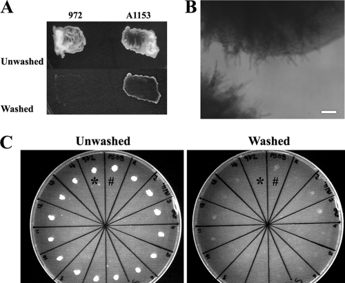
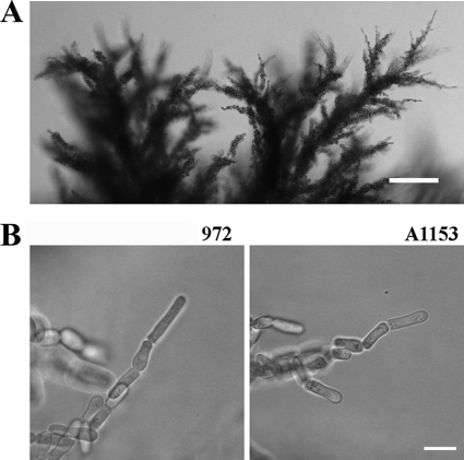
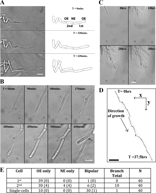
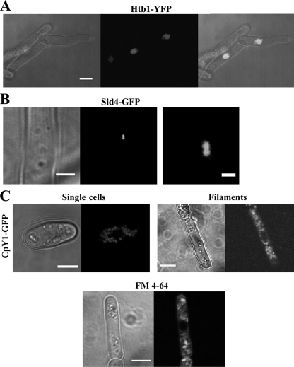
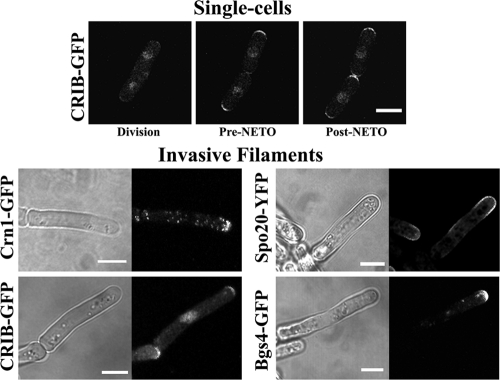
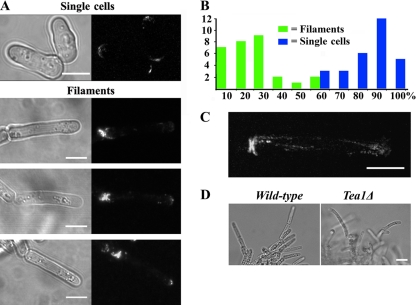
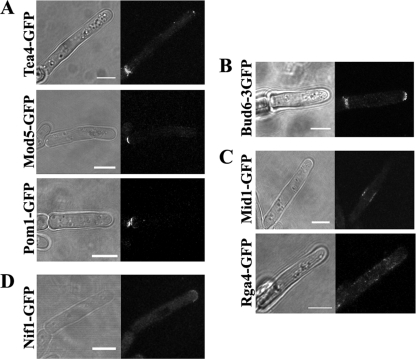
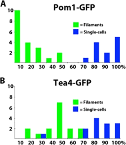
The organization and control of polarized growth through the cell cycle of Schizosaccharomyces pombe, a single-celled eukaryote, have been studied extensively. We have investigated the changes in these processes when S. pombe differentiates to form multicellular invasive mycelia and have found striking alterations to the behavior of some of the key regulatory proteins. Cells at the tips of invading filaments are considerably more elongated than cells growing singly and grow at one pole only. The filament tip follows a strict direction of growth through multiple cell cycles. A group of proteins involved in the growth process and actin regulation, comprising Spo20, Bgs4, activated Cdc42, and Crn1, are all concentrated at the growing tip, unlike their distribution at both ends of single cells. In contrast, several proteins implicated in microtubule-dependent organization of growth, including Tea1, Tea4, Mod5, and Pom1, all show the opposite effect and are relatively depleted at the growing end and enriched at the nongrowing end, although Tea1 appears to continue to be delivered to both ends. A third group acting at different stages of the cell cycle, including Bud6, Rga4, and Mid1, localize similarly in filaments and single cells, while Nif1 shows a reciprocal localization to Pom1.


Figures

Fig. 1.
Fig. 1.
Strain A1153 invades more efficiently than strain 972. (A) Strains 972 and A1153 were plated on LNB medium at high density, and after 4 days, the surface cells were removed by washing. (B) Strain A1153 forms invasive filaments that extend beyond the immediate area of the surface colony. Bar, 50 μm. (C) Mating of strain A1153 with strain 972. Strains 972 (*), A1153 (#), and 14 progeny of a cross between the two (remaining segments) were incubated for 2 days. Strain A1153 had invaded more strongly than strain 972, while the progeny from the cross showed various levels of invasion intermediate between the two parental strains.
Fig. 2.
Fig. 2.
The filamentous morphologies of strains A1153 and 972 are similar. (A) Invasive structures formed by strain A1153 after several weeks. The structures are large and complex and resemble those formed by strain 972. Bar, 100 μm. (B) Filaments at single-cell resolution. After 3 days, invasive structures formed by both strains (strains A1153 and 972) were observed in agar slices. The average filament size at division was estimated by measuring the lengths of septated filament tips (Table 2). Bar, 10 μm.
Fig. 3.
Fig. 3.
Time-lapse microscopy of S. pombe filamentous growth. (A) Montage of time-lapse images with cell outlines traced on the right. Filament tip cells are elongated (Table 2), grow monopolarly throughout the cell cycle, and undergo complete cell division, and no structures that attach adjacent cells are obvious. “1st” and “2nd” refer to the cell at the tip of the filament and the cell directly behind it, and “OE” and “NE” refer to the old and new ends, respectively. (B) Branch formation. Following division, the daughter behind the filament tip can grow monopolarly from its “new” end. More frequently, these daughters grow monopolarly from the “old” end as in panel A. (C) Selected time points from the time-lapse microscopy in Movie S1 in the supplemental material. (D) The position of the filament at each 15-min time point was plotted from Movie S1 in the supplemental material. Through 37.5 h of growth, encompassing nine cell cycles, the filament maintains a stable direction of growth. The straight guidance line represents a distance of 79.5 μm. Bar, 10 μm. (E) Quantitation of the patterns of filamentous growth. From time-lapse microscopy of growing filaments, the 1st and 2nd cells were scored as growing monopolarly from the new or old ends or bipolarly from both. The number of branched cells (branched cells shown in panel B) that were found in each class is shown in parentheses. In some cases, adjacent cells separated to allow them to grow past each other (middle image in panel A). This was classed as bipolar growth without branching. Single cells growing on LNB medium were analyzed in parallel. Time (T) is shown in minutes or hours.
Fig. 4.
Fig. 4.
Cellular organelles in filamentous cells. (A) Filamentous cells stained with nuclear marker Htb1-YFP (YFP stands for yellow fluorescent protein) (strain JA1551). The right panel shows the transmission and fluorescence microscopy images superimposed. The extended filaments are mononucleate. Bar, 5 μm. (B) Filamentous cells stained with Sid4-GFP, a marker for the spindle pole body (SPB) (strain JA1576). The right panel is a higher magnification of the duplicated SPB seen in the left panel. Bars, 2 μm (left panel) and 0.5 μm (right panel). (C) Vacuole morphology in filamentous cells. Filamentous cells were stained with vacuole lumen marker CpY-GFP (strain JA1582) (top) or with vital dye FM 4-64 (bottom), which labels endocytic structures and vacuole membranes. The size of vacuoles appears more irregular in filaments, and they are absent from a region near the tip. All images are single planes. Bars, 5 μm.
Fig. 5.
Fig. 5.
Four factors required for growth are restricted to the filament tip. The indicated polarity factors are enriched at the growing end of the filaments (strains JA1523, JA1543, JA1577, and JA1545). Single cells are shown at division and before and after new-end take-off (pre-NETO and post-NETO, respectively). Fluorescent images are maximum-intensity stacks; light transmission images are single planes. Bars, 5 μm.
Fig. 6.
Fig. 6.
Tea1 abundance at the growing tip is reduced in filamentous cells. (A) Tea1-3GFP distribution in single cells and filaments (strain JA1489). In single cells, Tea1-3GFP is located equally at both tips. In filaments, it is enriched strongly at the nongrowing end. Bars, 5 μm. (B) Quantitation of Tea1-3GFP distribution. Maximum-intensity stacks of Tea1-3GFP fluorescence were collected for both singly growing and filamentous cells of strain JA1489. The graph shows the fluorescence intensity of Tea1-3GFP at the growing end of the filament as a percentage of the intensity at the nongrowing end, plotted as 10% bins. For single cells, the fluorescence from the end with weaker intensity is plotted relative to the intensity from the other end. A total of 29 cells or filaments were examined. (C) Time-lapse imaging of Tea1-3GFP delivery to the tip of a filamentous cell in strain JA1489 (see Movie S2 in the supplemental material). Tea1-3GFP appears to be delivered equally to both ends of the filament. The image shows a maximum-intensity stack taken at 10-s intervals over 480 s. Bar, 5 μm. (D) A strain deleted for Tea1 (strain JA1567) produces filaments that are bent, resulting in aberrant mycelial morphology compared to that of the wild type (see Movie S3 in the supplemental material). Bar, 10 μm.
Fig. 7.
Fig. 7.
Distribution of seven other growth and polarity factors in filamentous cells. (A) Tea4-GFP, Mod5-GFP, and Pom1-GFP (from strains JA1542, JA1578, and JA1500, respectively) are enriched strongly only at the nongrowing ends of filamentous cells. (B) Bud6-3GFP (strain JA1476) is localized at both ends of the filament as in single cells. (C) Mid1-GFP and Rga4-GFP (from strains JA1501 and JA1527, respectively) exhibit a pattern of localization in filamentous cells similar to that found in single cells. (D) Nif1-GFP (strain JA1556) is enriched strongly at the growing end of the filamentous cell. Bars, 5 μm.
Fig. 8.
Fig. 8.
Quantitation of Pom1-GFP (A) and Tea4-GFP (B) distribution, measured as for Tea1 (Fig. 6). Tea4 shows a degree of polarization similar to that for Tea1, while Pom1 appears to be more stringently polarized.

Similar articles

Cited by

References

    1. Amoah-Buahin E., Bone N., Armstrong J. 2005. Hyphal growth in the fission yeast Schizosaccharomyces pombe. Eukaryot. Cell 4:1287–1297 - PMC - PubMed
    1. Bahler J., Pringle J. R. 1998. Pom1p, a fission yeast protein kinase that provides positional information for both polarized growth and cytokinesis. Genes Dev. 12:1356–1370 - PMC - PubMed
    1. Cortes J. C. G., Konomi M., Martins I. M., Munoz J., Moreno M. B., Osumi M., Duran A., Ribas J. C. 2007. The (1,3)beta-D-glucan synthase subunit Bgs1p is responsible for the fission yeast primary septum formation. Mol. Microbiol. 65:201–217 - PubMed
    1. Ding R., West R. R., Morphew M., Oakley B. R., McIntosh J. R. 1997. The spindle pole body of Schizosaccharomyces pombe enters and leaves the nuclear envelope as the cell cycle proceeds. Mol. Biol. Cell 8:1461–1479 - PMC - PubMed
    1. Dodgson J., Avula H., Hoe K. L., Kim D. U., Park H. O., Hayles J., Armstrong J. 2009. Functional genomics of adhesion, invasion, and mycelial formation in Schizosaccharomyces pombe. Eukaryot. Cell 8:1298–1306 - PMC - PubMed

Publication types

MeSH terms

Substances