. 2011 Jul;45(1):111-9.
doi: 10.1165/rcmb.2009-0445OC. Epub 2010 Sep 24.

Anti-inflammatory effects of thiazolidinediones in human airway smooth muscle cells

Affiliations

Anti-inflammatory effects of thiazolidinediones in human airway smooth muscle cells

Ming Zhu et al. Am J Respir Cell Mol Biol. 2011 Jul.

Abstract

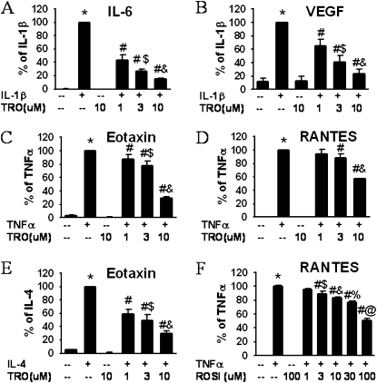
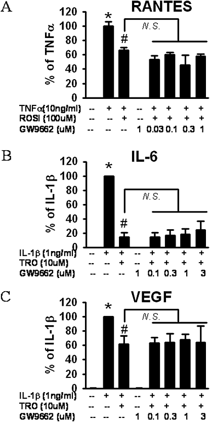
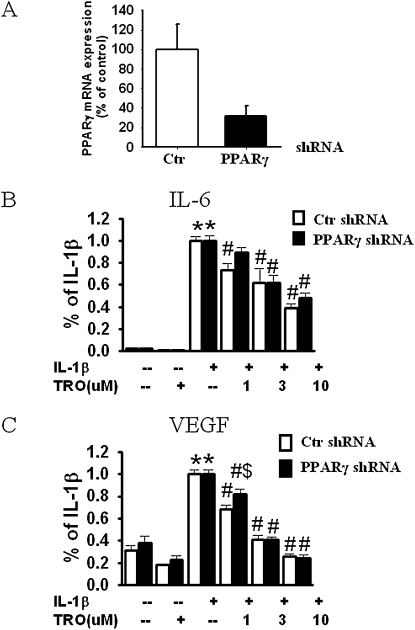
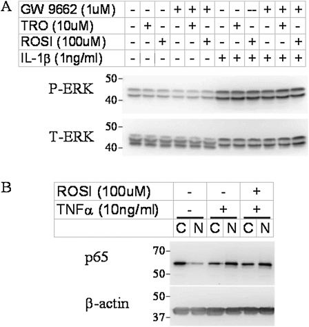
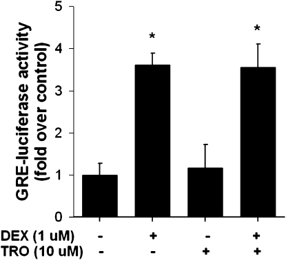
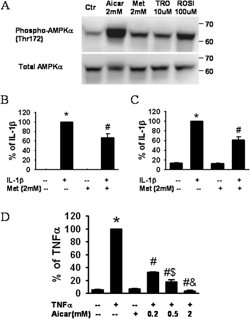
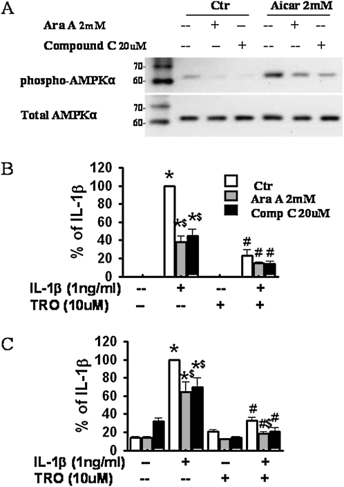
Airway smooth muscle (ASM) cells have been reported to contribute to the inflammation of asthma. Because the thiazolidinediones (TZDs) exert anti-inflammatory effects, we examined the effects of troglitazone and rosiglitazone on the release of inflammatory moieties from cultured human ASM cells. Troglitazone dose-dependently reduced the IL-1β-induced release of IL-6 and vascular endothelial growth factor, the TNF-α-induced release of eotaxin and regulated on activation, normal T expressed and secreted (RANTES), and the IL-4-induced release of eotaxin. Rosiglitazone also inhibited the TNF-α-stimulated release of RANTES. Although TZDs are known to activate peroxisome proliferator-activated receptor-γ (PPARγ), these anti-inflammatory effects were not affected by a specific PPARγ inhibitor (GW 9662) or by the knockdown of PPARγ using short hairpin RNA. Troglitazone and rosiglitazone each caused the activation of adenosine monophosphate-activated protein kinase (AMPK), as detected by Western blotting using a phospho-AMPK antibody. The anti-inflammatory effects of TZDs were largely mimicked by the AMPK activators, 5-amino-4-imidazolecarboxamide ribose (AICAR) and metformin. However, the AMPK inhibitors, Ara A and Compound C, were not effective in preventing the anti-inflammatory effects of troglitazone or rosiglitzone, suggesting that the effects of these TZDs are likely not mediated through the activation of AMPK. These data indicate that TZDs inhibit the release of a variety of inflammatory mediators from human ASM cells, suggesting that they may be useful in the treatment of asthma, and the data also indicate that the effects of TZDs are not mediated by PPARγ or AMPK.


Figures

Figure 1.
Figure 1.
Thiazolidinediones (TZDs) dose-dependently reduced the release of inflammatory mediators from human airway smooth muscle (HASM) cells. ELISA results are from HASM cell supernatants collected 24 hours after stimulation with IL-1β (1 ng/ml) (A, B), TNF-α (10 ng/ml) (C, D, F), or IL-4 (3 ng/ml) (E). Cells were treated with troglitazone (TRO) (A–E) or rosiglitazone (ROSI) (F). Results are presented as mean ± SE of data from cells from three or four donors, and under each condition, experiments were performed in triplicate or quadruplicate. Values were normalized to cell numbers in each well. *P < 0.05 compared with untreated cells. #P < 0.05 compared with cells treated with stimulator (IL-1β, IL-4, or TNF-α) alone. $P < 0.05 compared with cells treated with IL-1β or TNF-α plus 1 μM or 3 μM troglitazone, respectively. %,@P < 0.05 compared with cells treated with TNF-α plus 10 μM or 30 μM rosiglitazone, respectively. RANTES, regulated on activation, normal T expressed and secreted; VEGF, vascular endothelial growth factor.
Figure 2.
Figure 2.
Effect of the proliferator-activated receptor-γ (PPARγ) inhibitor, GW 9662, on the inhibition of RANTES (A), IL-6 (B), or VEGF (C) release from HASM cells by TZDs, according to ELISA. Increasing doses of GW 9662 were administered 2 hours before stimulation with TNF-α (A) or IL-1β (B, C). Troglitazone (TRO) (A, B) or rosiglitazone (ROSI) (C) were added 1 hour before the administration of cytokines. Results are presented as mean ± SE of data from three donors; each studied in duplicate, and normalized to cell number in each well. *P < 0.05 compared with untreated cells. #P < 0.05 compared with cells treated with IL-1β or TNF-α alone. N.S., no significance.
Figure 3.
Figure 3.
PPARγ knockdown does not prevent the anti-inflammatory effects of TZDs in HASM cells. (A) PPARγ mRNA level in HASM cells that were transduced with lentivirus transduction particles containing nontarget short hairpin RNA (shRNA) (control; Ctr) or PPARγ-target shRNA. Successfully transduced cells were selected by puromycin, expanded, and used in this study. (B, C) Pooled results of all four constructs from two donor cell lines. Results are normalized to maximal release of cytokine with IL-1β stimulation from the same donor treated with control shRNA, and are presented as mean ± SE. *P < 0.05 compared with untreated groups. #P < 0.05 compared with IL-1β–stimulated groups with the same shRNA treatment. $P < 0.05 compared with control shRNA with the same treatment.
Figure 4.
Figure 4.
Representative Western blot shows the effects of TZDs on IL-1β–induced phosphorylation of extracellular signal-regulated protein kinase (ERK) (A) and on TNF-α–induced activation of nuclear factor (NF)–κB (B) in HASM cells. Cells were treated for 15 minutes with IL-1β (A) or for 30 minutes with TNF-α (B). TZDs were administered 30 minutes before cytokines, and GW 9662 was administered 30 minutes before TZDs, if used. Twenty micrograms (A) or 10 μg (B) of protein per sample were separated on 10% SDS-PAGE gel. The activation of NF-κB was assessed by p65 translocation from the cytosolic (C) to the nuclear (N) fraction. P-ERK: phosphorylated ERK; T-ERK: total ERK.
Figure 5.
Figure 5.
Luciferase expression in untreated HASM cells and in cells treated with dexamethasone (DEX, 1 μM), troglitazone (TRO, 10 μM), or DEX + TRO for 1 hour and transfected with glucocorticoid-responsive element–dependent (GRE) luciferase. Results are presented as mean ± SE of data from three cell wells per treatment. Experiments were performed on cell lines derived from two different donors. *P < 0.05 versus no treatment.
Figure 6.
Figure 6.
(A) Western blot of HASM cell lysates shows phosphorylated and total adenosine monophosphate-activated kinase (AMPK) after incubation with the AMPK agonists, 5-aminoimidazole-4-carboxamide ribonucleoside (AICAR) and metformin (Met), or the TZDs troglitazone (TRO) and rosiglitazone (ROSI). Concentrations of (B) IL-6, (C) VEGF, and (D) eotaxin in HASM cell supernatants collected 24 hours after stimulation with IL-1β (1 ng/ml) (B, C) or TNF-α (10 ng/ml) (D), in the presence or absence of metformin (B, C) or AICAR (D). Results are presented as mean ± SE of data from cells of three donors, each assayed in duplicate and normalized to the cell number in each well. *P < 0.05 compared with untreated cells. #P < 0.05 compared with cells stimulated with IL-1β or TNF-α alone. $P < 0.05 compared with cells stimulated with TNF-α plus 0.2 mM or 0.5 mM AICAR, respectively.
Figure 7.
Figure 7.
Effects of the AMPK inhibitors, Ara A and Compound C, on the release of proinflammatory mediators from HASM cells. (A) Representative Western blot shows phosphorylated and total AMPK in lysates of control HASM cells and cells treated with the AMPK agonist, AICAR, in the presence or absence of AMPK inhibitors Ara A and Compound C. (B, C) ELISA results for IL-6 (B) and VEGF (C) in supernatants of HASM cells treated with IL-1β and/or troglitazone (TRO), in the presence or absence of Ara A and Compound C. Results are presented as mean ± SE of data from cells of three donors, each studied in duplicate. *P < 0.05 compared with untreated groups. #P < 0.05 compared with IL-1β–treated groups with the same AMPK inhibitors (Ara A and Compound C). $P < 0.05 compared with cells with IL-1β alone or IL-1β plus 10 μM TRO treatment.

Similar articles

Cited by

References

    1. Barnes PJ. The cytokine network in asthma and chronic obstructive pulmonary disease. J Clin Invest 2008;118:3546–3556. - PMC - PubMed
    1. Afshar R, Medoff BD, Luster AD. Allergic asthma: a tale of many T cells. Clin Exp Allergy 2008;38:1847–1857. - PubMed
    1. Bousquet J, Chanez P, Lacoste JY, Barneon G, Ghavanian N, Enander I, Venge P, Ahlstedt S, Simony-Lafontaine J, Godard P, et al. Eosinophilic inflammation in asthma. N Engl J Med 1990;323:1033–1039. - PubMed
    1. Ghaffar O, Hamid Q, Renzi PM, Allakhverdi Z, Molet S, Hogg JC, Shore SA, Luster AD, Lamkhioued B. Constitutive and cytokine-stimulated expression of eotaxin by human airway smooth muscle cells. Am J Respir Crit Care Med 1999;159:1933–1942. - PubMed
    1. Faffe DS, Flynt L, Mellema M, Moore PE, Silverman ES, Subramaniam V, Jones MR, Mizgerd JP, Whitehead T, Imrich A, et al. Oncostatin M causes eotaxin-1 release from airway smooth muscle: synergy with IL-4 and IL-13. J Allergy Clin Immunol 2005;115:514–520. - PubMed

Publication types

MeSH terms

Substances