. 2010 Sep;22(9):3053-65.
doi: 10.1105/tpc.110.074351. Epub 2010 Sep 24.

The Arabidopsis exocyst complex is involved in cytokinesis and cell plate maturation

Affiliations

The Arabidopsis exocyst complex is involved in cytokinesis and cell plate maturation

Matyás Fendrych et al. Plant Cell. 2010 Sep.

Abstract

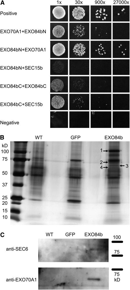
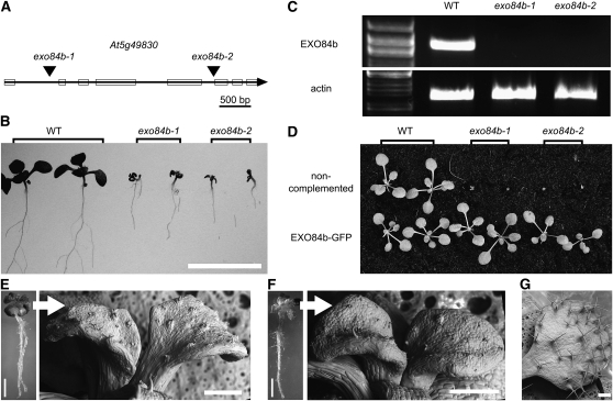
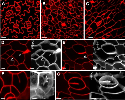
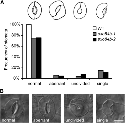
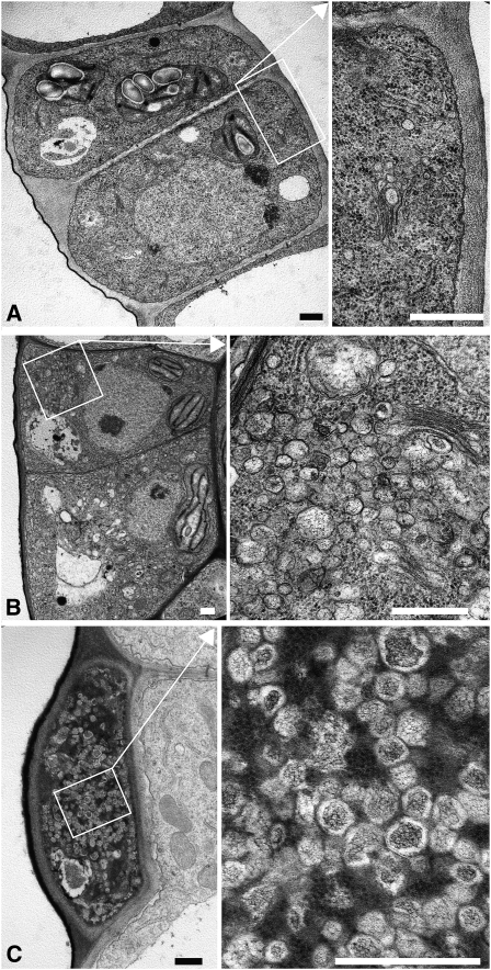
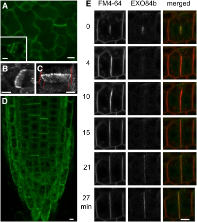
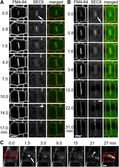
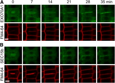
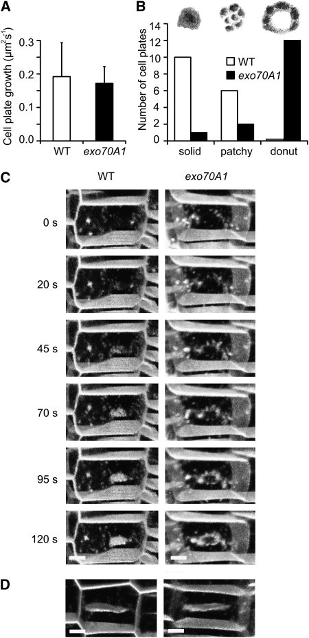
Cell reproduction is a complex process involving whole cell structures and machineries in space and time, resulting in regulated distribution of endomembranes, organelles, and genomes between daughter cells. Secretory pathways supported by the activity of the Golgi apparatus play a crucial role in cytokinesis in plants. From the onset of phragmoplast initiation to the maturation of the cell plate, delivery of secretory vesicles is necessary to sustain successful daughter cell separation. Tethering of secretory vesicles at the plasma membrane is mediated by the evolutionarily conserved octameric exocyst complex. Using proteomic and cytologic approaches, we show that EXO84b is a subunit of the plant exocyst. Arabidopsis thaliana mutants for EXO84b are severely dwarfed and have compromised leaf epidermal cell and guard cell division. During cytokinesis, green fluorescent protein-tagged exocyst subunits SEC6, SEC8, SEC15b, EXO70A1, and EXO84b exhibit distinctive localization maxima at cell plate initiation and cell plate maturation, stages with a high demand for vesicle fusion. Finally, we present data indicating a defect in cell plate assembly in the exo70A1 mutant. We conclude that the exocyst complex is involved in secretory processes during cytokinesis in Arabidopsis cells, notably in cell plate initiation, cell plate maturation, and formation of new primary cell wall.


Figures

Figure 1.
Figure 1.
EXO84b Interacts with Other Exocyst Subunits. (A) Pairwise interactions of EXO84b N- and C-terminal fragments in the yeast two-hybrid system with exocyst subunits EXO70A1 and SEC15b. The strength of interactions is demonstrated by a dilution series (indicated on the top). Binding and activation domains are denoted on the left and right, respectively. (B) and (C) Coimmunoprecipitation of exocyst subunits with GFP-tagged EXO84b. (B) EXO84b, SEC15b, SEC10, and EXO70A1 (1, 2, 3, and 4, respectively) were identified in the immunoprecipitate by mass spectrometry. (C) SEC6 and EXO70A1 were detected by immunoblotting. Approximate protein molecular mass in kilodaltons is indicated, and the samples were loaded in the same ratio as in (B).
Figure 2.
Figure 2.
Mutations in EXO84b Lead to a Severe Growth Phenotype. (A) EXO84b gene structure with T-DNA insertion positions indicated. (B) Wild-type, exo84b-1, and exo84b-2 seedlings 10 d after germination. Bar = 1 cm. (C) RT-PCR of the full-length EXO84b. The ACTIN7 control was amplified from the same cDNA. (D) Comparison of uncomplemented (top row) and exo84b-1 and exo84b-2 plants complemented by EXO84b-GFP expressed under 35S and EXO84b promoter (bottom row), respectively. (E) to (G) exo84b-1 (E) and exo84b-2 (F) mutants have minute true leaves with defective trichomes. On the left, 10-d-old seedlings (bar = 1 mm); on the right, detail of the true leaves is shown (environmental scanning electron microscopy). Wild-type leaf is shown in (G). Bars = 200 μm.
Figure 3.
Figure 3.
exo84b Mutants Have Cell Wall Stubs. (A) to (C) FM4-64–stained abaxial 3rd leaf epidermis of wild-type (A), exo84b-1 (B), and exo84b-2 (C) seedlings. (D) to (G) FM4-64–stained abaxial leaf epidermal cells of exo84b-1 mutant plants. A single confocal section is shown in red (left) with corresponding three-dimensional reconstruction from serial confocal sections shown in white with the rotation indicated (arrow). Epidermal cells contained short cell wall stubs (asterisk) (D), long unfinished cell walls (triangle) ([D] and [E]), and aberrant cell plates (F). (G) Imperfectly separated guard cells with a developed stomatal pore. After the three-dimensional reconstruction, unfinished cell plate connected with the stomatal pore is evident. Bars = 10 μm.
Figure 4.
Figure 4.
Stomata Morphology in exo84b Mutants. (A) Frequency of stomata sorted by their morphology in leaves of 12-d-old wild type (white), exo84b-1 (gray), and exo84b-2 (black); n > 200 for each genotype. The category “normal” also includes developing stomata. (B) Most abundant stomata types: normal stomata, aberrant stomata with highly asymmetric guard cells, stomata with incompletely divided guard cells, and stomata consisting of a single guard cell. Bar = 10 μm.
Figure 5.
Figure 5.
exo84b Mutants Accumulate Vesicles. Transmission electron microscopy sections of epidermal cells of the wild type (A) and exo84b mutants ([B] and [C]). A proportion of exo84b mutant cells accumulated vesicles (B). A dead cell loaded with vesicles in the epidermis of exo84b-2 mutant plant (C). The boxed areas are shown in higher magnification on the right, with the positions of the boxes indicated with arrows. Bars = 0.5 μm.
Figure 6.
Figure 6.
EXO84b-GFP Fusion Protein Localizes to the Cell Plate and Postcytokinetic Wall. (A) Abaxial epidermis of a developing leaf shows strong EXO84b-GFP signal at the cross walls. Recently divided guard cells with EXO84b-GFP–labeled ventral walls are shown in the inset. (B) and (C) Three-dimensional reconstructions of CLSM sections of the leaf (B) and root (C) cross walls. In (B), the 35S-EXO84b-GFP particles are visible. In (C), perpendicular cell walls in respect to the newly formed cell walls are indicated with red lines. (D) EXO84b-GFP labels postcytokinetic walls in the Arabidopsis root. (E) Time series of a root growing cell plate stained with FM4-64. EXO84b-GFP signal localized to the newly emerged cell plate. In the cell plate growth phase, the signal intensity decreases and reappears with the cell plate insertion into the mother plasma membrane. In (A), (D), and (E), EXO84b was expressed under EXO84b promoter and in (B) and (C), under 35S promoter. In the confocal laser scanning microscopy sections, bars = 5 μm.
Figure 7.
Figure 7.
SEC6 and SEC8 Localize to the Cell Plate and Postcytokinetic Wall. (A) and (B) Time series showing SEC6-GFP (A) and GFP-SEC8 (B) localization during cytokinesis of root meristem cells stained with FM4-64. Arrow denotes strong association of both proteins with the new cell plate. Arrowhead points to cell plate insertion site labeled by SEC6-GFP and GFP-SEC8. Single confocal laser scanning microscopy sections. (C) The initial flash of SEC6-GFP signal (see text) is marked with an arrow. Arrowhead points to SEC6-GFP postcytokinetic wall signal. The position of the cell is indicated with red lines, three-dimensional reconstruction of CLSM sections taken in a time series, slanted backwards. Bars = 5 μm.
Figure 8.
Figure 8.
Localization Dynamics of GFP-EXO70A1 and GFP-SEC15b during Cytokinesis. GFP-EXO70A1 and GFP-SEC15b localize to the nascent cell plate; later, the signal reappears at the time of cell plate insertion to the mother wall and expands along the whole area of the postcytokinetic wall. Cell membranes were stained by FM4-64. Single confocal laser scanning microscopy sections are displayed. Bars = 5 μm.
Figure 9.
Figure 9.
Analysis of exo70A1 Mutant Cytokinesis. (A) Mean cell plate growth rate of the wild type and exo70A1; error bars represent sd. (B) Classification and count of solid, patchy, and donut-shaped cell plate types as sketched at the top found in wild-type and exo70A1 mutant cells. (C) Initial phases of cytokinesis of the wild type (left) and exo70A1 (right) in cells stained with FM4-64; donut-shaped cell plate is obvious in the mutant. (D) In later phases, the wild-type and exo70A1 cell plates were not distinguishable. Bars = 5 μm.

References

    1. Abramoff M.D., Magelhaes P.J., Ram S.J. (2004). Image Processing with ImageJ. Biophotonics Int. 11: 36–42
    1. Buschmann H., Chan J., Sanchez-Pulido L., Andrade-Navarro M.A., Doonan J.H., Lloyd C.W. (2006). Microtubule-associated AIR9 recognizes the cortical division site at preprophase and cell-plate insertion. Curr. Biol. 16: 1938–1943 - PubMed
    1. Chiu W., Niwa Y., Zeng W., Hirano T., Kobayashi H., Sheen J. (1996). Engineered GFP as a vital reporter in plants. Curr. Biol. 6: 325–330 - PubMed
    1. Chow C.M., Neto H., Foucart C., Moore I. (2008). Rab-A2 and Rab-A3 GTPases define a trans-Golgi endosomal membrane domain in Arabidopsis that contributes substantially to the cell plate. Plant Cell 20: 101–123 - PMC - PubMed
    1. Clough S.J., Bent A.F. (1998). Floral dip: A simplified method for Agrobacterium-mediated transformation of Arabidopsis thaliana. Plant J. 16: 735–743 - PubMed

Publication types

Substances