. 2010 Dec 15;19(24):4861-70.
doi: 10.1093/hmg/ddq419. Epub 2010 Sep 24.

Mitofusin 1 and mitofusin 2 are ubiquitinated in a PINK1/parkin-dependent manner upon induction of mitophagy

Affiliations

Mitofusin 1 and mitofusin 2 are ubiquitinated in a PINK1/parkin-dependent manner upon induction of mitophagy

Matthew E Gegg et al. Hum Mol Genet. .

Erratum in

  • Hum Mol Genet. 2013 Apr 15;22(8):1697

Abstract

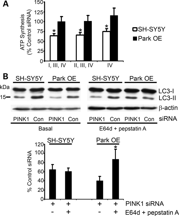
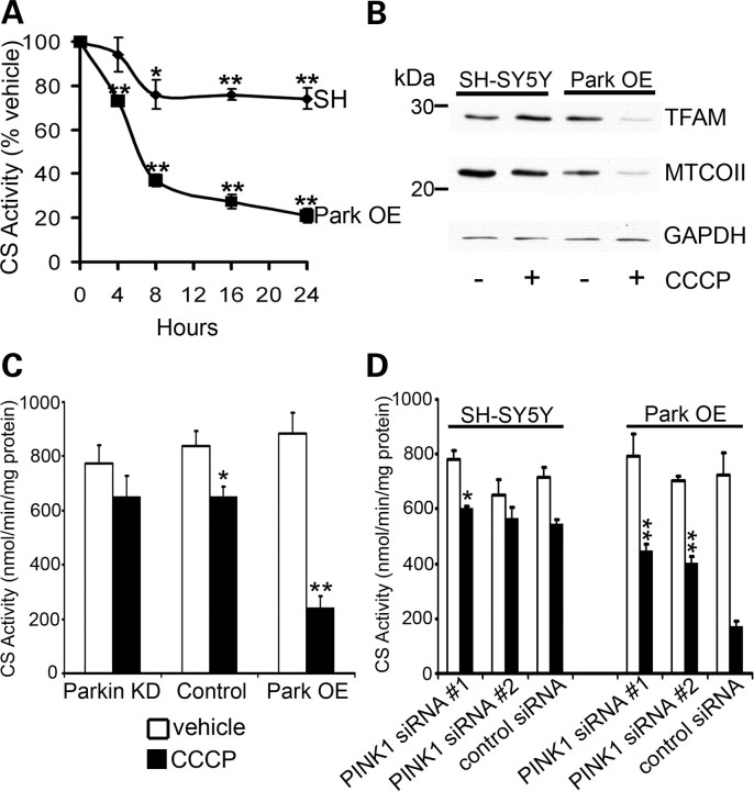
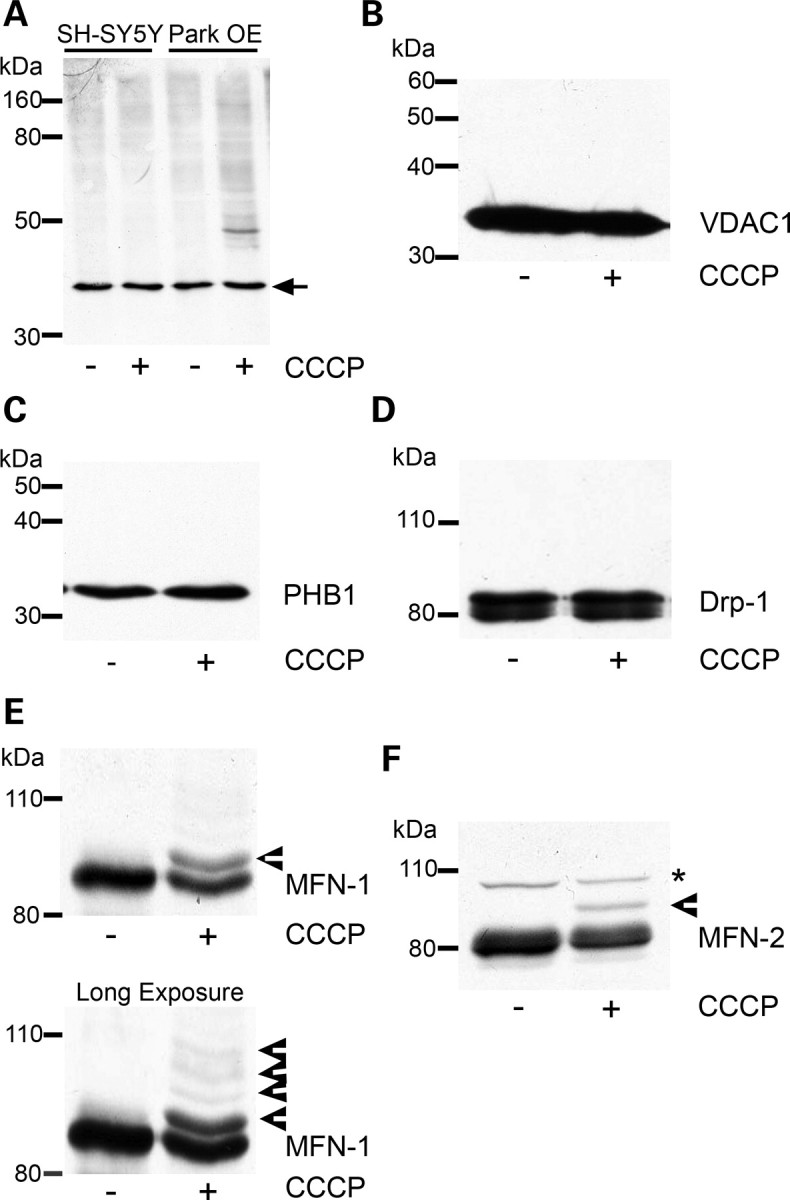
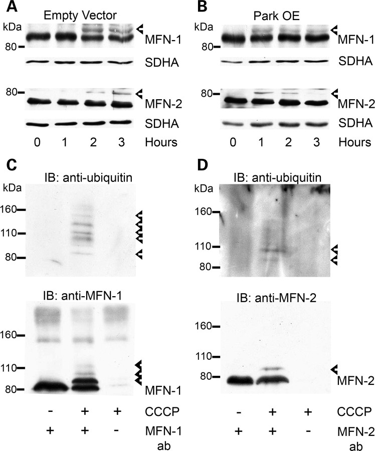
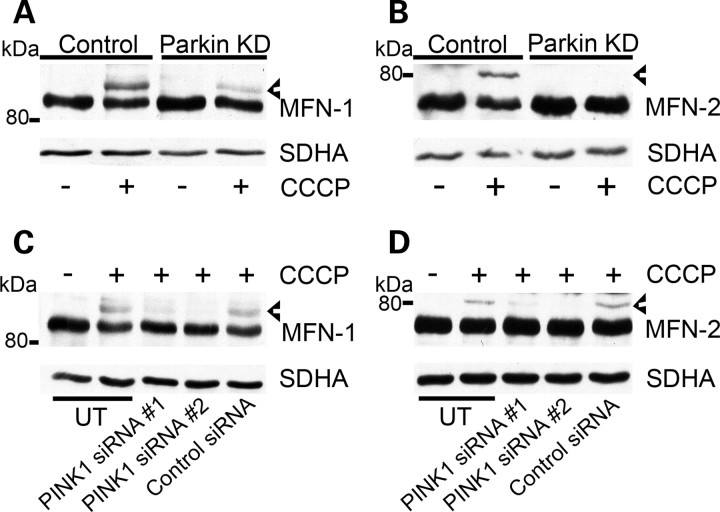
Mitochondrial dysfunction and perturbed degradation of proteins have been implicated in Parkinson's disease (PD) pathogenesis. Mutations in the Parkin and PINK1 genes are a cause of familial PD. PINK1 is a putative kinase associated with mitochondria, and loss of PINK1 expression leads to mitochondrial dysfunction, which increases with time. Parkin is suggested to be downstream of PINK1 and also mediates the removal of damaged mitochondria by macroautophagy (mitophagy). We investigated whether mitochondrial dysfunction in dopaminergic SH-SY5Y cells following decreased PINK1 expression by RNAi may in part be due to the inhibition of mitophagy. Reduced flux through the macroautophagy pathway was found to be coincident with the inhibition of ATP synthesis following 12 days of PINK1 silencing. Overexpression of parkin in these cells restored both autophagic flux and ATP synthesis. Overexpression and RNAi studies also indicated that PINK1 and parkin were required for mitophagy following CCCP-induced mitochondrial damage. The ubiquitination of several mitochondrial proteins, including mitofusin 1 and mitofusin 2, were detected within 3 h of CCCP treatment. These post-translational modifications were reduced following the silencing of parkin or PINK1. The ubiquitination of mitochondrial proteins appears to identify mitochondria for degradation and facilitate mitophagy. PINK1 and parkin are thus required for the removal of damaged mitochondria in dopaminergic cells, and inhibition of this pathway may lead to the accumulation of defective mitochondria which may contribute to PD pathogenesis.


Figures

Figure 1.
Figure 1.
ATP synthesis and autophagy flux after 12 days of PINK1 silencing. SH-SY5Y cells or Park OE cells were transfected with PINK1 siRNA #2 or control siRNA for 12 days. (A) Cells were harvested by trypsinization, permeabilized with digitonin and incubated with glutamate + malate (complexes I, III, IV), succinate + rotenone (complexes II, III, IV) or ascorbate + TMPD (complex IV) at 37°C to measure ATP synthesis. ATP was measured by a luciferase assay and normalized to cell number. Data are expressed as % control siRNA ATP synthesis (mean ± SEM; n = 5). *P < 0.05 versus control siRNA. (B) Cell lysates were prepared from untreated cells (basal) or cells treated with the lysosomal inhibitors E64d and pepstatin A (both 10 μg/ml) for 3 h. Western blots were probed with LC3 antibody and equal protein loading was assessed using an antibody against β-actin. The density of bands was measured and the LC3-II/LC3-I ratio was normalized to β-actin. Data are expressed as % of control siRNA-treated cell lines under basal conditions or in the presence of lysosomal inhibitors (n = 5). *P < 0.05 versus ParkSH + siRNA under basal conditions.
Figure 2.
Figure 2.
CCCP-induced mitophagy requires parkin and PINK1 expression. (A) SH-SY5Y cells containing an empty vector (SH) or Park OE cells were treated with vehicle [0.05% (v/v) ethanol] or 10 μm CCCP, lysed and CS activity assessed. Data are expressed as % CS activity of vehicle-treated cells (mean ± SEM; n = 5). *P < 0.05 versus vehicle and **P < 0.01 versus vehicle. (B) Cells were treated with vehicle or CCCP for 24 h, lysed and mitochondrial protein expression (TFAM, MTCOII) assessed by western blot. Equal loading was assessed by probing for GAPDH. Blot representative of four separate experiments. (C) Normal SH-SY5Y cells (control), parkin KD cells or Park OE cells were treated with vehicle or CCCP for 16 h, lysed and CS activity measured. Data are expressed as nmol/min/mg protein (mean ± SEM; n = 5). *P < 0.05 versus vehicle and **P < 0.01 versus vehicle. (D) SH-SY5Y cells or Park OE cells were treated with PINK1 siRNA or control siRNA for 72 h. For the last 16 h, cells were treated with vehicle or CCCP. CS activity was then measured. Data are expressed as nmol/min/mg protein (mean ± SEM; n = 5). *P < 0.05 versus control siRNA + CCCP and **P < 0.01 versus control siRNA + CCCP.
Figure 3.
Figure 3.
Ubiquitination of mitochondrial proteins following CCCP treatment. (A) SH-SY5Y cells containing an empty vector or Park OE cells were treated with vehicle [0.05% (v/v) ethanol] or 10 µm CCCP for 3 h, mitochondria isolated and a western blot performed. The western blot was probed with an antibody against ubiquitin. Equal loading was determined by probing for the 39 kDa subunit of complex I (arrow). Blot representative of four separate experiments. (B) Park OE cells were treated with CCCP for 3 h, mitochondria isolated and probed for VDAC1 or (C) prohibitin (PHB1) by western blot. (D) Park OE cells were treated with CCCP for 3 h and cell lysates probed for Drp1 by western blot. (E) Mitochondria isolated from Park OE cells treated with vehicle or CCCP for 3 h were probed for MFN-1 or (F) MFN-2 by western blot. Post-translationally modified proteins are indicated by arrowheads. Non-specific band is denoted by an asterisk. Blots are representative of at least four separate experiments.
Figure 4.
Figure 4.
Ubiquitination of MFN-1 and MFN-2 following CCCP treatment. (A) SH-SY5Y cells containing an empty vector or (B) Park OE cells were treated with 10 µm CCCP for 0–3 h, cell lysates prepared and MFN-1 and MFN-2 post-translational modifications assessed by western blot using antibodies against MFN-1 and MFN-2. Blots were re-probed with an antibody against the mitochondrial protein SDHA, to assess mitochondrial content in each lysate. (C) Mitochondria were isolated from SH-SY5Y cells containing an empty vector treated with vehicle or 10 µm CCCP for 2 h. Endogenous MFN-1 was immunoprecipitated from mitochondrial fractions and ubiquitinated species detected by western blotting. The blot was then re-probed with MFN-1 antibody. (D) MFN-2 was immunoprecipitated from mitochondrial fractions and ubiquitination detected as above. Blots are representative of at least three separate experiments. Ubiquitinated species are denoted by arrowheads.
Figure 5.
Figure 5.
Parkin and PINK1 are required for ubiquitination of MFN-1 and MFN-2. (A) Control or Parkin KD cells were treated with vehicle or CCCP for 2 h, cell lysates prepared and MFN-1 or (B) MFN-2 ubiquitination assessed by western blot using antibodies against MFN-1 and MFN-2. (C) SH-SY5Y cells were untransfected (UT), transfected with PINK1 siRNA #1 or PINK1 siRNA #2 or transfected with control siRNA for 72 h. Untransfected cells and siRNA transfected cells were then treated with 10 µm CCCP for 2 h, cell lysates prepared and separated by western blot. Blots were probed with an antibody against MFN-1 or (D) MFN-2. Blots were re-probed with an antibody against SDHA for mitochondrial content. Blots are representative of at least three separate experiments. Ubiquitinated species are denoted by arrowheads.

References

    1. Schapira A.H., Tolosa E. Molecular and clinical prodrome of Parkinson disease: implications for treatment. Nat. Rev. Neurol. 2010;6:309–317. doi:10.1038/nrneurol.2010.52. - DOI - PubMed
    1. Cuervo A.M., Wong E.S., Martinez-Vicente M. Protein degradation, aggregation, and misfolding. Mov. Disord. 2010;25:S49–S54. doi:10.1002/mds.22718. - DOI - PubMed
    1. Schapira A.H., Cooper J.M., Dexter D., Jenner P., Clark J.B., Marsden C.D. Mitochondrial complex I deficiency in Parkinson's disease. Lancet. 1989;1:1269. doi:10.1016/S0140-6736(89)92366-0. - DOI - PubMed
    1. Schapira A.H.V. Mitochondria in the aetiology and pathogenesis of Parkinson's disease. Lancet Neurol. 2008;7:97–109. doi:10.1016/S1474-4422(07)70327-7. - DOI - PubMed
    1. Betarbet R., Sherer T.B., MacKenzie G., Garcia-Osuna M., Panov A.V., Greenamyre J.T. Chronic systemic pesticide exposure reproduces features of Parkinson's disease. Nat. Neurosci. 2000;3:1301–1306. doi:10.1038/81834. - DOI - PubMed

Publication types

MeSH terms