Nodal and extranodal plasmacytomas expressing immunoglobulin a: an indolent lymphoproliferative disorder with a low risk of clinical progression
- PMID: 20871216
- PMCID: PMC2947321
- DOI: 10.1097/PAS.0b013e3181f17d0d
Nodal and extranodal plasmacytomas expressing immunoglobulin a: an indolent lymphoproliferative disorder with a low risk of clinical progression
Abstract
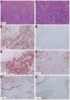
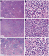
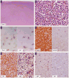
Plasmacytomas expressing immunoglobulin A are rare and not well characterized. In this study, 9 cases of IgA-positive plasmacytoma presenting in lymph node and 3 in extranodal sites were analyzed by morphology, immunohistochemistry, and polymerase chain reaction examination of immunoglobulin heavy and κ light chain genes. Laboratory features were correlated with clinical findings. There were 7 males and 5 females; age range was 10 to 66 years (median, 32 y). Six of the patients were younger than 30 years of age, 5 of whom had nodal disease. About 67% (6 of 9) of the patients with nodal disease had evidence of immune system dysfunction, including human immunodeficiency virus infection, T-cell deficiency, autoantibodies, arthritis, Sjögren syndrome, and decreased B cells. An IgA M-spike was detected in 6 of 11 cases, and the M-protein was nearly always less than 30 g/L. All patients had an indolent clinical course without progression to plasma cell myeloma. Histologically, nodal IgA plasmacytomas showed an interfollicular or diffuse pattern of plasma cell infiltration. The plasma cells were generally of mature Marschalko type with little or mild pleomorphism and exclusive expression of monotypic IgA. There was an equal expression of κ and λ light chains (ratio 6:6). Clonality was showed in 9 of 12 cases: by polymerase chain reaction in 7 cases, by cytogenetic analysis in 1 case, and by immunofixation in 1 case. Clonality did not correlate with pattern of lymph node infiltration. Our results suggest that IgA plasmacytomas may represent a distinct form of extramedullary plasmacytoma characterized by younger age at presentation, frequent lymph node involvement, and low risk of progression to plasma cell myeloma.
Figures


Similar articles
-
Primary plasmacytoma of lymph nodes.Hum Pathol. 1997 Sep;28(9):1083-90. doi: 10.1016/s0046-8177(97)90063-0. Hum Pathol. 1997. PMID: 9308734
-
Lack of expression of surface immunoglobulin light chains in B-cell non-Hodgkin lymphomas.Am J Clin Pathol. 2000 Mar;113(3):399-405. doi: 10.1309/28ED-MM0T-DT3B-MT4P. Am J Clin Pathol. 2000. PMID: 10705821
-
Improved clonality analysis based on immunoglobulin kappa locus for canine cutaneous plasmacytoma.Vet Immunol Immunopathol. 2019 Sep;215:109903. doi: 10.1016/j.vetimm.2019.109903. Epub 2019 Jul 24. Vet Immunol Immunopathol. 2019. PMID: 31420067
-
Primary extramedullary plasmacytoma with diffuse lymph node involvement: a case report and review of the literature.J Med Case Rep. 2019 May 22;13(1):153. doi: 10.1186/s13256-019-2087-7. J Med Case Rep. 2019. PMID: 31113466 Free PMC article. Review.
-
Primary extramedullary plasmacytoma of the lung.Intern Med. 1992 Dec;31(12):1396-400. doi: 10.2169/internalmedicine.31.1396. Intern Med. 1992. PMID: 1300176 Review.
Cited by
-
Plasma cell neoplasms and related entities-evolution in diagnosis and classification.Virchows Arch. 2023 Jan;482(1):163-177. doi: 10.1007/s00428-022-03431-3. Epub 2022 Nov 21. Virchows Arch. 2023. PMID: 36414803 Free PMC article. Review.
-
Loss-of-function of the protein kinase C δ (PKCδ) causes a B-cell lymphoproliferative syndrome in humans.Blood. 2013 Apr 18;121(16):3117-25. doi: 10.1182/blood-2012-12-469544. Epub 2013 Feb 21. Blood. 2013. PMID: 23430113 Free PMC article.
-
Extramedullary plasmacytoma in children is a genetically distinct localized neoplasia curable by surgical resection.Blood Adv. 2025 Aug 12;9(15):3909-3918. doi: 10.1182/bloodadvances.2025016596. Blood Adv. 2025. PMID: 40493885 Free PMC article.
-
Coexisting and clonally identical classic hodgkin lymphoma and nodular lymphocyte predominant hodgkin lymphoma.Am J Surg Pathol. 2011 May;35(5):767-72. doi: 10.1097/PAS.0b013e3182147f91. Am J Surg Pathol. 2011. PMID: 21490448 Free PMC article.
-
[B-cell neoplasms with plasmacellular and plasmablastic differentiation].Pathologe. 2013 May;34(3):198-209. doi: 10.1007/s00292-013-1743-8. Pathologe. 2013. PMID: 23462793 Review. German.
References
-
- Alexiou C, Kau RJ, Dietzfelbinger H, et al. Extramedullary plasmacytoma: tumor occurrence and therapeutic concepts. Cancer. 1999;85:2305–2314. - PubMed
-
- Bartl R, Frisch B, Fateh-Moghadam A, et al. Histologic classification and staging of multiple myeloma. A retrospective and prospective study of 674 cases. Am J Clin Pathol. 1987;87:342–355. - PubMed
-
- Bernstein SC, Perez-Atayde AR, Weinstein HJ. Multiple myeloma in a child. Cancer. 1985;56:2143–2147. - PubMed
-
- Bila J, Suvajdzic N, Elezovic I, et al. Systemic lupus Erythematosus and IgA multiple myeloma: a rare association? Medical oncology (Northwood, London, England) 2007;24:445–448. - PubMed
-
- Campo E, Pileri SA, Jaffe ES, et al. Nodal marginal zone lymphoma. In: Swerdlow SH, Campo E, Harris NL, et al., editors. WHO Classification of Tumours of Haematopoietic and Lymphoid Tissues. Lyon: International Agency for Research on Cancer; 2008. pp. 218–219.
Publication types
MeSH terms
Substances
Grants and funding
LinkOut - more resources
Full Text Sources
Miscellaneous

