. 2010 Nov 3;29(21):3723-32.
doi: 10.1038/emboj.2010.238. Epub 2010 Sep 24.

TopBP1 functions with 53BP1 in the G1 DNA damage checkpoint

Affiliations

TopBP1 functions with 53BP1 in the G1 DNA damage checkpoint

Rachele Cescutti et al. EMBO J. .

Abstract

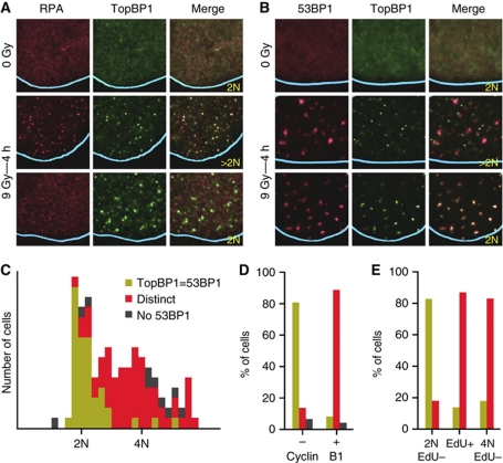
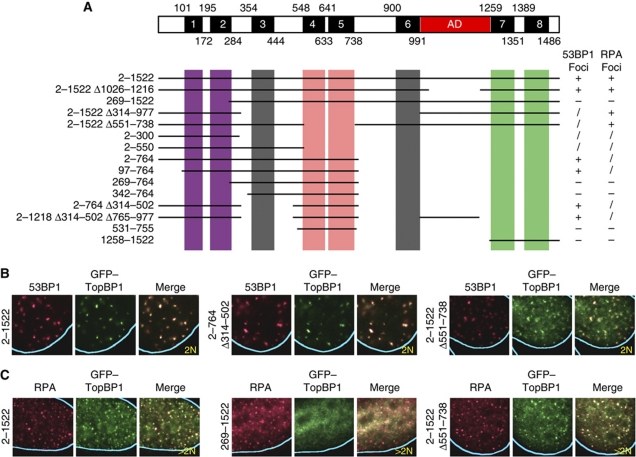
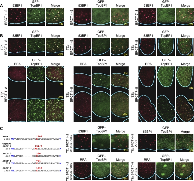
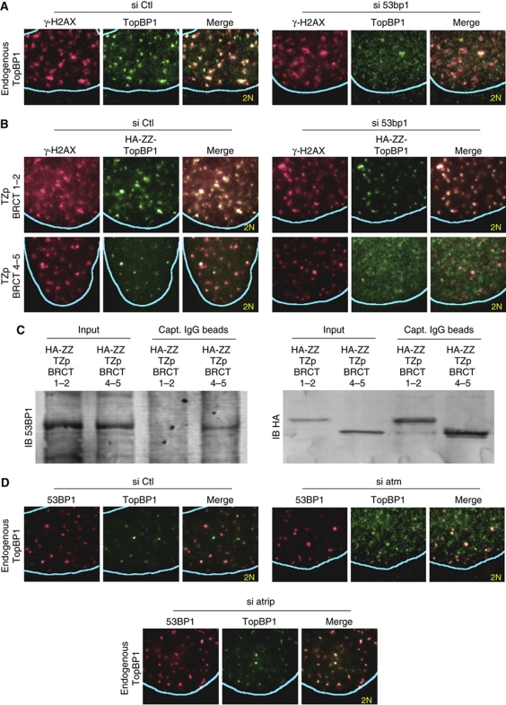
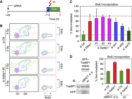
TopBP1 is a checkpoint protein that colocalizes with ATR at sites of DNA replication stress. In this study, we show that TopBP1 also colocalizes with 53BP1 at sites of DNA double-strand breaks (DSBs), but only in the G1-phase of the cell cycle. Recruitment of TopBP1 to sites of DNA replication stress was dependent on BRCT domains 1-2 and 7-8, whereas recruitment to sites of DNA DSBs was dependent on BRCT domains 1-2 and 4-5. The BRCT domains 4-5 interacted with 53BP1 and recruitment of TopBP1 to sites of DNA DSBs in G1 was dependent on 53BP1. As TopBP1 contains a domain important for ATR activation, we examined whether it contributes to the G1 cell cycle checkpoint. By monitoring the entry of irradiated G1 cells into S-phase, we observed a checkpoint defect after siRNA-mediated depletion of TopBP1, 53BP1 or ATM. Thus, TopBP1 may mediate the checkpoint function of 53BP1 in G1.


Conflict of interest statement

The authors declare that they have no conflict of interest.

Figures

Figure 1
Figure 1
Endogenous TopBP1 colocalizes with either RPA or 53BP1 in irradiated cells depending on the phase of the cell cycle. (A, B) Localization of endogenous TopBP1, RPA and 53BP1 in U2OS cells 4 h after exposure to IR (9 Gy) or after mock-irradiation (0 Gy), as determined by immunofluorescence. Because the IR-induced RPA foci are much smaller than the 53BP1 foci, only part of the cell nucleus is shown with the blue lines indicating the periphery of the nucleus. In this and all other figures the DNA content of the cells was quantified on the basis of the intensity of DAPI staining over the entire nucleus and shown as 2 or >2N; the DAPI staining intensities of the adjacent cells were used for calibration. (C) Histogram plot of the behaviour of TopBP1 according to genomic DNA content. Endogenous TopBP1 and 53BP1 IR-induced foci were visualized in U2OS cells 4 h after exposure to IR (9 Gy). The behaviour of TopBP1 was scored as: TopBP1=53BP1, colocalized TopBP1 and 53BP1 IR-induced foci; distinct, non-colocalizing TopBP1 and 53BP1 foci; No 53BP1, no 53BP1 foci. (D, E) Colocalization of TopBP1 and 53BP1 IR-induced foci according to (D) Cyclin B1 staining or genomic DNA content and (E) EdU incorporation. The behaviour of TopBP1 is presented using the same colouring scheme as in (C).
Figure 2
Figure 2
Mapping the domains that mediate recruitment of TopBP1 to IR-induced 53BP1 or RPA foci. (A) Diagram of human TopBP1 showing the BRCT domains, numbered 1–8, and the ATR activation domain (AD) and summary of the properties of the tested TopBP1 deletion mutants. The mutants were scored according to efficiency of focus formation: +, wild type; /, foci formed, but a significant amount of TopBP1 remained in the nucleoplasm; −, no foci. (B, C) Examples of the localization of selected GFP-tagged TopBP1 mutants in U2OS cells 4 h after exposure to IR (9 Gy). The numbers refer to the residues present in the TopBP1 mutants. Δ, deletion.
Figure 3
Figure 3
Different pairs of TopBP1 BRCT domains have distinct properties for recruitment to sites of DNA damage. (A) Recruitment of BRCT domains 1–2 (residues 2–300), 4–5 (residues 531–755) and 7–8 (residues 1258–1522) as monomers to sites of DNA damage. Domains 1–2 formed IR-induced foci with reduced efficiency compared with full-length TopBP1; domains 4–5 and 7–8 did not form IR-induced foci. In this and all subsequent panels, U2OS cells were examined 4 h after irradiation (9 Gy). (B) Recruitment of BRCT domains 1–2 (residues 2–300), 4–5 (residues 531–755) and 7–8 (residues 1258–1522) as tetramers (TZp) to sites of DNA damage. Tetramers of domains 1–2 colocalized with both 53BP1 and RPA IR-induced foci. Tetramers of domains 4–5 colocalized only with 53BP1 foci and then only in G1 cells. Tetramers of domains 7–8 colocalized only with RPA foci. (C) Potential phosphate-binding lysines in BRCT domains 1, 2, 5 and 7 of human TopBP1 and effect of amino acid substitutions targeting these lysines on recruitment of oligomerized TopBP1 BRCT domains to IR-induced foci. The putative phosphate-binding lysines are coloured red; other conserved BRCT domain residues are coloured blue. The corresponding sequence of BRCA1 is shown for reference.
Figure 4
Figure 4
TopBP1 recruitment to sites of DNA DSBs is 53BP1- and ATM-dependent. (A) Immunofluorescence analysis of U2OS cells for endogenous TopBP1 and phosphorylated histone H2AX (γ-H2AX) 4 h after irradiation (9 Gy). The cells were transfected with control (Ctl) siRNA or siRNA targeting 53bp1. (B) Immunofluorescence analysis of U2OS cells stably expressing haemagglutinin peptide and protein A-tagged (HA-ZZ) oligomerized (TZp) TopBP1 BRCT domains 1–2 (residues 2–300) or 4–5 (residues 531–755). The cells were transfected with control (Ctl) siRNA or siRNA targeting 53bp1 and were monitored for localization of γ-H2AX and the stably expressed TopBP1 proteins 4 h after irradiation (9 Gy). (C) Co-precipitation of oligomerized TopBP1 BRCT domains 4–5 with endogenous 53BP1. U2OS cells stably expressing HA-ZZ-tagged tetramers (TZp) of RCT domains 1–2 (residues 2–300) or 4–5 (residues 531–755) were used to prepare nuclear extracts (Input), which were then incubated with IgG-coated beads. HA-ZZ–TZp–TopBP1 fusion proteins and endogenous 53BP1 bound to the beads were detected by immunoblotting (IB) using anti-HA and anti-53BP1 antibodies, respectively. (D) Immunofluorescence analysis of U2OS cells for endogenous 53BP1 and TopBP1 4 h after irradiation (9 Gy). The cells were transfected with control (Ctl) siRNA or siRNA targeting atm or atrip.
Figure 5
Figure 5
TopBP1 and 53BP1 are required for the G1 DNA damage checkpoint. (A) Design of the G1 checkpoint assay. U2OS cells were transfected with siRNA. After 48 h, the cells were pulsed with EdU (E) for 1 h and then exposed to 2 Gy IR. After irradiation, the cells were incubated with BrdU and nocodazole for 7 h and then examined by flow cytometry. (B) Representative examples of flow cytometry data. Cells having G1 DNA content and staining negatively for EdU (highlighted by the purple circles in the left panels) were plotted on the basis of BrdU versus EdU incorporation (right panels). (C) Statistical analysis of the results of the G1 checkpoint assay performed in quadruplicate. For each sample, the fraction of BrdU-positive cells over the total number of G1/Edu-negative cells was calculated; then the ratio was determined of the fraction for the irradiated cells divided by the fraction for the non-irradiated cells. A ratio of 100% indicates no arrest in G1 after irradiation (marked by a red horizontal line). The green horizontal line indicates the ratio for the cells transfected with control (Ctl) siRNA. Means and s.d. values are shown. (D) A TopBP1 mutant lacking BRCT domains 4–5 is defective in the G1 checkpoint assay. U2OS cells stably transfected with plasmids expressing GFP-tagged wild-type (wt) TopBP1 or a TopBP1 mutant lacking residues 551–738 were treated with control (Ctl) siRNA or siRNA targeting the endogenous topbp1 gene (the 3′ untranslated region; si topbp1 #2) and then subjected to the G1 checkpoint assay. Expression of the endogenous and ectopically expressed TopBP1 proteins was monitored by immunoblotting (IB) with antibodies against TopBP1 and GFP. −; non-transfected cells.

References

    1. Abraham RT (2001) Cell cycle checkpoint signaling through the ATM and ATR kinases. Genes Dev 15: 2177–2196 - PubMed
    1. Anderson L, Henderson C, Adachi Y (2001) Phosphorylation and rapid relocalization of 53BP1 to nuclear foci upon DNA damage. Mol Cell Biol 21: 1719–1729 - PMC - PubMed
    1. Ball HL, Cortez D (2005) ATRIP oligomerization is required for ATR-dependent checkpoint signaling. J Biol Chem 280: 31390–31396 - PMC - PubMed
    1. Bothmer A, Robbiani DF, Feldhahn N, Gazumyan A, Nussenzweig A, Nussenzweig MC (2010) 53BP1 regulates DNA resection and the choice between classical and alternative end joining during class switch recombination. J Exp Med 207: 855–865 - PMC - PubMed
    1. Bouwman P, Aly A, Escandell JM, Pieterse M, Bartkova J, van der Gulden H, Hiddingh S, Thanasoula M, Kulkarni A, Yang Q, Haffty BG, Tommiska J, Blomqvist C, Drapkin R, Adams DJ, Nevanlinna H, Bartek J, Tarsounas M, Ganesan S, Jonkers J (2010) 53BP1 loss rescues BRCA1 deficiency and is associated with triple-negative and BRCA-mutated breast cancers. Nat Struct Mol Biol 17: 688–695 - PMC - PubMed

Publication types

MeSH terms