

Tissue is alive: New technologies are needed to address the problems of protein biomarker pre-analytical variability
- PMID: 20871745
- PMCID: PMC2944287
- DOI: 10.1002/prca.200800001
Tissue is alive: New technologies are needed to address the problems of protein biomarker pre-analytical variability
Abstract
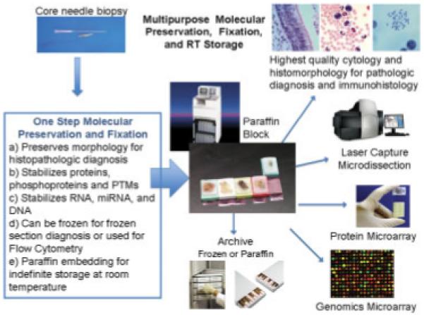
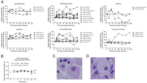
Instability of tissue protein biomarkers is a critical issue for molecular profiling. Pre-analytical variables during tissue procurement, such as time delays during which the tissue remains stored at room temperature, can cause significant variability and bias in downstream molecular analysis. Living tissue, ex vivo, goes through a defined stage of reactive changes that begin with oxidative, hypoxic and metabolic stress, and culminate in apoptosis. Depending on the delay time ex vivo, and reactive stage, protein biomarkers, such as signal pathway phosphoproteins will be elevated or suppressed in a manner which does not represent the biomarker levels at the time of excision. Proteomic data documenting reactive tissue protein changes post collection indicate the need to recognize and address tissue stability, preservation of post-translational modifications, and preservation of morphologic features for molecular analysis. Based on the analysis of phosphoproteins, one of the most labile tissue protein biomarkers, we set forth tissue procurement guidelines for clinical research. We propose technical solutions for (i) assessing the state of protein analyte preservation and specimen quality via identification of a panel of natural proteins (surrogate stability markers), and (ii) using multi-purpose fixative solution designed to stabilize, preserve and maintain proteins, nucleic acids, and tissue architecture.
Figures
References
-
- Wilker E, Lu J, Rho O, Carbajal S, et al. Role of PI3K/Akt signaling in insulin-like growth factor-1 (IGF-1) skin tumor promotion. Mol. Carcinog. 2005;44:137–145. - PubMed
-
- Sun SY, Rosenberg LM, Wang X, Zhou Z, et al. Activation of Akt and eIF4E survival pathways by rapamycin-mediated mammalian target of rapamycin inhibition. Cancer Res. 2005;65:7052–7058. - PubMed
-
- Shah A, Swain WA, Richardson D, Edwards J, et al. Phospho-akt expression is associated with a favorable outcome in non-small cell lung cancer. Clin. Cancer Res. 2005;11:2930–2936. - PubMed
-
- Calvo E, Rowinsky EK. Effect of epidermal growth factor receptor mutations on the response to epidermal growth factor receptor tyrosine kinase inhibitors: target-based populations for target-based drugs. Clin. Lung Cancer. 2004;6:S35–S42. - PubMed
-
- Huang S, Armstrong EA, Benavente S, Chinnaiyan P, et al. Dual-agent molecular targeting of the epidermal growth factor receptor (EGFR): combining anti-EGFR antibody with tyrosine kinase inhibitor. Cancer Res. 2004;64:5355–5362. - PubMed
Grants and funding
LinkOut - more resources
Full Text Sources
Other Literature Sources
