

Adult retinal stem cells revisited
- PMID: 20871757
- PMCID: PMC2945004
- DOI: 10.2174/1874364101004010030
Adult retinal stem cells revisited
Abstract
Recent advances in retinal stem cell research have raised the possibility that these cells have the potential to be used to repair or regenerate diseased retina. Various cell sources for replacement of retinal neurons have been identified, including embryonic stem cells, the adult ciliary epithelium, adult Müller stem cells and induced pluripotent stem cells (iPS). However, the true stem cell nature of the ciliary epithelium and its possible application in cell therapies has now been questioned, leaving other cell sources to be carefully examined as potential candidates for such therapies. The need for identification of the ontogenetic state of grafted stem cells in order to achieve their successful integration into the murine retina has been recognized. However, it is not known whether the same requirements may apply to achieve transplant cell integration into the adult human eye. In addition, the existence of natural barriers for stem cell transplantation, including microglial accumulation and abnormal extracellular matrix deposition have been demonstrated, suggesting that several obstacles need to be overcome before such therapies may be implemented. This review addresses recent scientific developments in the field and discusses various strategies that may be potentially used to design cell based therapies to treat human retinal disease.
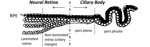
Keywords: Human retina; Müller stem cells; adult stem cells; ciliary marginal zone; regeneration; transplantation..
Figures
References
-
- Evans MJ, Kaufman MH. Establishment in culture of pluripotential cells from mouse embryos. Nature. 1981;292(5819):154–6. - PubMed
-
- Bibel M, Richter J, Schrenk K, et al. Differentiation of mouse embryonic stem cells into a defined neuronal lineage. Nat Neurosci. 2004;7(9):1003–9. - PubMed
-
- Hirano M, Yamamoto A, Yoshimura N, et al. Generation of structures formed by lens and retinal cells differentiating from embryonic stem cells. Dev Dyn. 2003;228(4):664–71. - PubMed
-
- Hemmati-Brivanlou A, Melton D. Vertebrate embryonic cells will become nerve cells unless told otherwise. Cell. 1997;88(1):13–7. - PubMed
Grants and funding
LinkOut - more resources
Full Text Sources
Other Literature Sources