

PPARα in Obesity: Sex Difference and Estrogen Involvement
- PMID: 20871824
- PMCID: PMC2943125
- DOI: 10.1155/2010/584296
PPARα in Obesity: Sex Difference and Estrogen Involvement
Abstract
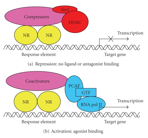
Peroxisome proliferator-activated receptor α (PPARα) is a member of the steroid hormone receptor superfamily and is well known to act as the molecular target for lipid-lowering drugs of the fibrate family. At the molecular level, PPARα regulates the transcription of a number of genes critical for lipid and lipoprotein metabolism. PPARα activators are further shown to reduce body weight gain and adiposity, at least in part, due to the increase of hepatic fatty acid oxidation and the decrease in levels of circulating triglycerides responsible for adipose cell hypertrophy and hyperplasia. However, these effects of the PPARα ligand fenofibrate on obesity are regulated with sexual dimorphism and seem to be influenced by the presence of functioning ovaries, suggesting the involvement of ovarian steroids in the control of obesity by PPARα. In female ovariectomized mice, 17β-estradiol inhibits the actions of fenofibrate on obesity through its suppressive effects on the expression of PPARα target genes, and these processes may be mediated by inhibiting the coactivator recruitment of PPARα. Thus, it is likely that PPARα functions on obesity may be enhanced in estrogen-deficient states.
Figures


Similar articles
-
The role of PPARalpha in lipid metabolism and obesity: focusing on the effects of estrogen on PPARalpha actions.Pharmacol Res. 2009 Sep;60(3):151-9. doi: 10.1016/j.phrs.2009.02.004. Epub 2009 Feb 14. Pharmacol Res. 2009. PMID: 19646654 Review.
-
Inhibition of the actions of peroxisome proliferator-activated receptor alpha on obesity by estrogen.Obesity (Silver Spring). 2007 Jun;15(6):1430-40. doi: 10.1038/oby.2007.171. Obesity (Silver Spring). 2007. PMID: 17557980
-
Peroxisome proliferator-activated receptor alpha-isoform deficiency leads to progressive dyslipidemia with sexually dimorphic obesity and steatosis.J Biol Chem. 1998 Nov 6;273(45):29577-85. doi: 10.1074/jbc.273.45.29577. J Biol Chem. 1998. PMID: 9792666
-
Peroxisome proliferator-activated receptor alpha is not rate-limiting for the lipoprotein-lowering action of fish oil.J Biol Chem. 2001 Feb 16;276(7):4634-9. doi: 10.1074/jbc.M008809200. Epub 2000 Oct 24. J Biol Chem. 2001. PMID: 11050100
-
Regulation of lipid and lipoprotein metabolism by PPAR activators.Clin Chem Lab Med. 2000 Jan;38(1):3-11. doi: 10.1515/CCLM.2000.002. Clin Chem Lab Med. 2000. PMID: 10774955 Review.
Cited by
-
Associations between dietary patterns and gene expression profiles of healthy men and women: a cross-sectional study.Nutr J. 2013 Feb 12;12:24. doi: 10.1186/1475-2891-12-24. Nutr J. 2013. PMID: 23398686 Free PMC article.
-
Coenzyme A Restriction as a Factor Underlying Pre-Eclampsia with Polycystic Ovary Syndrome as a Risk Factor.Int J Mol Sci. 2022 Mar 3;23(5):2785. doi: 10.3390/ijms23052785. Int J Mol Sci. 2022. PMID: 35269927 Free PMC article.
-
Peroxisome proliferator-activated receptor α, a potential therapeutic target for alcoholic liver disease.World J Gastroenterol. 2014 Jul 7;20(25):8055-60. doi: 10.3748/wjg.v20.i25.8055. World J Gastroenterol. 2014. PMID: 25009377 Free PMC article. Review.
-
Genistein as a nature-derived PPAR agonist in adipogenesis and weight gain.Eur J Nutr. 2015 Apr;54(3):489-91. doi: 10.1007/s00394-015-0848-7. Eur J Nutr. 2015. PMID: 25662822 No abstract available.
-
Overview of Ethnobotanical-Pharmacological Studies Carried Out on Medicinal Plants from the Serra da Estrela Natural Park: Focus on Their Antidiabetic Potential.Pharmaceutics. 2024 Mar 25;16(4):454. doi: 10.3390/pharmaceutics16040454. Pharmaceutics. 2024. PMID: 38675115 Free PMC article. Review.
References
-
- Issemann I, Green S. Activation of a member of the steroid hormone receptor superfamily by peroxisome proliferators. Nature. 1990;347(6294):645–650. - PubMed
-
- Beck F, Plummer S, Senior PV, Byrne S, Green S, Brammar WJ. The ontogeny of peroxisome-proliferator-activated receptor gene expression in the mouse and rat. Proceedings of the Royal Society B. 1992;247(1319):83–87. - PubMed
-
- Braissant O, Foufelle F, Scotto C, Dauça M, Wahli W. Differential expression of peroxisome proliferator-activated receptors (PPARs): tissue distribution of PPAR-α, -β, and -γ in the adult rat. Endocrinology. 1996;137(1):354–366. - PubMed
-
- Aoyama T, Peters JM, Iritani N, et al. Altered constitutive expression of fatty acid-metabolizing enzymes in mice lacking the peroxisome proliferator-activated receptor α (PPARα) Journal of Biological Chemistry. 1998;273(10):5678–5684. - PubMed
-
- Auwerx J, Schoonjans K, Fruchart J-C, Staels B. Transcriptional control of triglyceride metabolism: fibrates and fatty acids change the expression of the LPL and apo C-III genes by activating the nuclear receptor PPAR. Atherosclerosis. 1996;124, supplement:S29–S37. - PubMed
LinkOut - more resources
Full Text Sources
