

Acinetobacter baumannii in Localised Cutaneous Mycobacteriosis in Falcons
- PMID: 20871867
- PMCID: PMC2943107
- DOI: 10.4061/2010/321797
Acinetobacter baumannii in Localised Cutaneous Mycobacteriosis in Falcons
Abstract
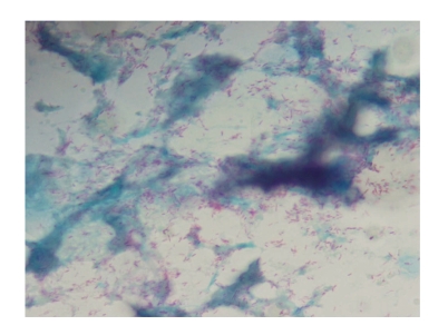
Between May 2007 and April 2009, 29 falcons with identically localized, yellowish discolored cutaneous lesions in the thigh and lateral body wall region were presented at Abu Dhabi Falcon Hospital. Out of 18 falcons integrated in this study, 16 tested positive to Mycobacterium. avium complex. The 2 negative falcons tested positive in the Mycobacterium genus PCR. Moreover, 1 falcon tested positive to M. avium. paratuberculosis in tissue samples by PCR. In all cases, blood and fecal samples tested negative. In the acid-fast stain, all samples showed the for mycobacteriosis typical rods. Moreover, in 13 samples Acinetobacter baumannii was detected by PCR and proven by DNA sequencing. Clinical features included highly elevated WBCs, heterophilia, lymphocytopenia, monocytosis, severe anemia and weight loss. A. baumannii, a gram-negative bacillus with the ability to integrate foreign DNA, has emerged as one of the major multidrug resistant bacteria. In veterinary medicine, it has so far been detected in dogs, cats, horses and wild birds. To the authors' knowledge, this is the first report of an A. baumannii infection in falcons and of a veterinary Mycobacterium-Acinetobacter coinfection.
Figures
Similar articles
-
Acinetobacter in veterinary medicine, with an emphasis on Acinetobacter baumannii.J Glob Antimicrob Resist. 2019 Mar;16:59-71. doi: 10.1016/j.jgar.2018.08.011. Epub 2018 Aug 23. J Glob Antimicrob Resist. 2019. PMID: 30144636 Review.
-
First detection of Cryptosporidium parvum in falcons (Falconiformes): Diagnosis, molecular sequencing, therapeutic trial and epidemiological assessment of a possible emerging disease in captive falcons.Vet Parasitol. 2018 Mar 15;252:167-172. doi: 10.1016/j.vetpar.2018.02.012. Epub 2018 Feb 9. Vet Parasitol. 2018. PMID: 29559142
-
Falcons From the United Arab Emirates Infected With Chlamydia psittaci/C abortus Intermediates Specified as Chlamydia buteonis by Polymerase Chain Reaction.J Avian Med Surg. 2021 Sep;35(3):333-340. doi: 10.1647/20-00050. J Avian Med Surg. 2021. PMID: 34677032
-
Multi-drug-resistant Acinetobacter calcoaceticus-Acinetobacter baumannii complex infection outbreak in dogs and cats in a veterinary hospital.J Small Anim Pract. 2016 Nov;57(11):617-625. doi: 10.1111/jsap.12555. Epub 2016 Oct 5. J Small Anim Pract. 2016. PMID: 27709647
-
Multidrug resistant Acinetobacter baumannii in veterinary medicine--emergence of an underestimated pathogen?Berl Munch Tierarztl Wochenschr. 2014 Nov-Dec;127(11-12):435-46. Berl Munch Tierarztl Wochenschr. 2014. PMID: 25872253 Review.
Cited by
-
Brain abscess and bronchopneumonia caused by Acinetobacter baumannii in a 2-year-old female sheep.Vet Q. 2018 Dec;38(1):67-71. doi: 10.1080/01652176.2018.1489165. Vet Q. 2018. PMID: 30375283 Free PMC article. No abstract available.
-
Acinetobacter baumannii from Samples of Commercially Reared Turkeys: Genomic Relationships, Antimicrobial and Biocide Susceptibility.Microorganisms. 2023 Mar 16;11(3):759. doi: 10.3390/microorganisms11030759. Microorganisms. 2023. PMID: 36985332 Free PMC article.
-
Low Occurrence of Acinetobacter baumannii in Gulls and Songbirds.Pol J Microbiol. 2020;69(1):1-6. doi: 10.33073/pjm-2020-011. Pol J Microbiol. 2020. PMID: 32162853 Free PMC article.
References
-
- Schreckenberger PC, Daneshvar MI, Weyant RS, Hollis DG. Acinetobacter, Achromobacter, Chryseobacterium, Moxarella, and other nonfermentive gram-negative rods. In: Murray PR, editor. Manual of Clinical Microbiology. 8th edition. Washington, DC, USA: ASM Press; 2003. pp. 749–779.
-
- Villers D, Espaze E, Coste-Burel M, et al. Nosocomial Acinetobacter baumannii infections: microbiological and clinical epidemiology. Annals of Internal Medicine. 1998;129(3):182–189. - PubMed
LinkOut - more resources
Full Text Sources
Other Literature Sources
Miscellaneous
