Translocator protein PET imaging for glial activation in multiple sclerosis
- PMID: 20872081
- PMCID: PMC3257858
- DOI: 10.1007/s11481-010-9243-6
Translocator protein PET imaging for glial activation in multiple sclerosis
Erratum in
- J Neuroimmune Pharmacol. 2011 Sep;6(3):434. Fujimura, Yota [added]; Richert, Nancy D [added]
Abstract
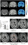
Glial activation in the setting of central nervous system inflammation is a key feature of the multiple sclerosis (MS) pathology. Monitoring glial activation in subjects with MS, therefore, has the potential to be informative with respect to disease activity. The translocator protein 18 kDa (TSPO) is a promising biomarker of glial activation that can be imaged by positron emission tomography (PET). To characterize the in vivo TSPO expression in MS, we analyzed brain PET scans in subjects with MS and healthy volunteers in an observational study using [(11)C]PBR28, a newly developed translocator protein-specific radioligand. The [(11)C]PBR28 PET showed altered compartmental distribution of TSPO in the MS brain compared to healthy volunteers (p = 0.019). Focal increases in [(11)C]PBR28 binding corresponded to areas of active inflammation as evidenced by significantly greater binding in regions of gadolinium contrast enhancement compared to contralateral normal-appearing white matter (p = 0.0039). Furthermore, increase in [(11)C]PBR28 binding preceded the appearance of contrast enhancement on magnetic resonance imaging in some lesions, suggesting a role for early glial activation in MS lesion formation. Global [(11)C]PBR28 binding showed correlation with disease duration (p = 0.041), but not with measures of clinical disability. These results further define TSPO as an informative marker of glial activation in MS.
Conflict of interest statement
Figures


Similar articles
-
Translocator protein 18 kDa (TSPO) expression in multiple sclerosis patients.J Neuroimmune Pharmacol. 2013 Mar;8(1):51-7. doi: 10.1007/s11481-012-9397-5. Epub 2012 Sep 6. J Neuroimmune Pharmacol. 2013. PMID: 22956240 Free PMC article.
-
A quantitative neuropathological assessment of translocator protein expression in multiple sclerosis.Brain. 2019 Nov 1;142(11):3440-3455. doi: 10.1093/brain/awz287. Brain. 2019. PMID: 31578541 Free PMC article.
-
Lower levels of the glial cell marker TSPO in drug-naive first-episode psychosis patients as measured using PET and [11C]PBR28.Mol Psychiatry. 2017 Jun;22(6):850-856. doi: 10.1038/mp.2016.247. Epub 2017 Feb 14. Mol Psychiatry. 2017. PMID: 28194003
-
Positron emission tomography imaging in multiple sclerosis-current status and future applications.Eur J Neurol. 2011 Feb;18(2):226-231. doi: 10.1111/j.1468-1331.2010.03154.x. Eur J Neurol. 2011. PMID: 20636368 Review.
-
Positron-emission tomography molecular imaging of glia and myelin in drug discovery for multiple sclerosis.Expert Opin Drug Discov. 2015 May;10(5):557-70. doi: 10.1517/17460441.2015.1032240. Epub 2015 Apr 5. Expert Opin Drug Discov. 2015. PMID: 25843125 Review.
Cited by
-
Neuroinflammatory component of gray matter pathology in multiple sclerosis.Ann Neurol. 2016 Nov;80(5):776-790. doi: 10.1002/ana.24791. Epub 2016 Oct 25. Ann Neurol. 2016. PMID: 27686563 Free PMC article.
-
Molecular imaging of multiple sclerosis: from the clinical demand to novel radiotracers.EJNMMI Radiopharm Chem. 2019 Apr 8;4(1):6. doi: 10.1186/s41181-019-0058-3. EJNMMI Radiopharm Chem. 2019. PMID: 31659498 Free PMC article. Review.
-
Potential Biomarkers Associated with Multiple Sclerosis Pathology.Int J Mol Sci. 2021 Sep 25;22(19):10323. doi: 10.3390/ijms221910323. Int J Mol Sci. 2021. PMID: 34638664 Free PMC article. Review.
-
18F-GE-180: a novel TSPO radiotracer compared to 11C-R-PK11195 in a preclinical model of stroke.Eur J Nucl Med Mol Imaging. 2015 Mar;42(3):503-11. doi: 10.1007/s00259-014-2939-8. Epub 2014 Oct 29. Eur J Nucl Med Mol Imaging. 2015. PMID: 25351507
-
Imaging the Influence of Red Blood Cell Docosahexaenoic Acid Status on the Expression of the 18 kDa Translocator Protein in the Brain: A [11C]PBR28 Positron Emission Tomography Study in Young Healthy Men.Biol Psychiatry Cogn Neurosci Neuroimaging. 2022 Oct;7(10):998-1006. doi: 10.1016/j.bpsc.2021.09.005. Epub 2021 Oct 2. Biol Psychiatry Cogn Neurosci Neuroimaging. 2022. PMID: 34607054 Free PMC article.
References
-
- Banati RB, Newcombe J, Gunn RN, et al. The peripheral benzodiazepine binding site in the brain in multiple sclerosis: quantitative in vivo imaging of microglia as a measure of disease activity. Brain. 2000;123(Pt 11):2321–2337. - PubMed
-
- Briard E, Zoghbi SS, Imaizumi M, et al. Synthesis and evaluation in monkey of two sensitive 11C-labeled aryloxyanilide ligands for imaging brain peripheral benzodiazepine receptors in vivo. J Med Chem. 2008;51:17–30. - PubMed
-
- Debruyne JC, Versijpt J, Van Laere KJ, et al. PET visualization of microglia in multiple sclerosis patients using [11C]PK11195. Eur J Neurol. 2003;10:257–264. - PubMed
Publication types
MeSH terms
Substances
Grants and funding
LinkOut - more resources
Full Text Sources
Medical

