. 2011 Jan;233(1):1-12.
doi: 10.1007/s00425-010-1275-x. Epub 2010 Sep 26.

Immunocytochemical determination of the subcellular distribution of ascorbate in plants

Affiliations

Immunocytochemical determination of the subcellular distribution of ascorbate in plants

Bernd Zechmann et al. Planta. 2011 Jan.

Abstract

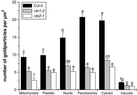
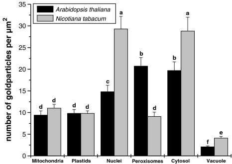
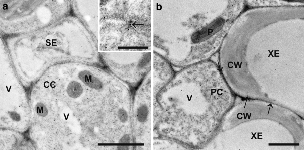
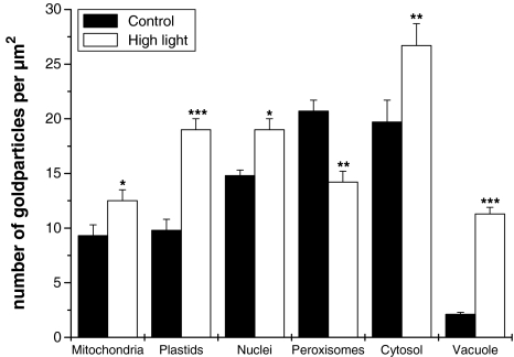
Ascorbate is an important antioxidant in plants and fulfills many functions related to plant defense, redox signaling and modulation of gene expression. We have analyzed the subcellular distribution of reduced and oxidized ascorbate in leaf cells of Arabidopsis thaliana and Nicotiana tabacum by high-resolution immuno electron microscopy. The accuracy and specificity of the applied method is supported by several observations. First, preadsorption of the ascorbate antisera with ascorbic acid or dehydroascorbic acid resulted in the reduction of the labeling to background levels. Second, the overall labeling density was reduced between 50 and 61% in the ascorbate-deficient Arabidopsis mutants vtc1-2 and vtc2-1, which correlated well with biochemical measurements. The highest ascorbate-specific labeling was detected in nuclei and the cytosol whereas the lowest levels were found in vacuoles. Intermediate labeling was observed in chloroplasts, mitochondria and peroxisomes. This method was used to determine the subcellular ascorbate distribution in leaf cells of plants exposed to high light intensity, a stress factor that is well known to cause an increase in cellular ascorbate concentration. High light intensities resulted in a strong increase in overall labeling density. Interestingly, the strongest compartment-specific increase was found in vacuoles (fourfold) and in plastids (twofold). Ascorbate-specific labeling was restricted to the matrix of mitochondria and to the stroma of chloroplasts in control plants but was also detected in the lumen of thylakoids after high light exposure. In summary, this study reveals an improved insight into the subcellular distribution of ascorbate in plants and the method can now be applied to determine compartment-specific changes in ascorbate in response to various stress situations.


Figures

Fig. 1
Fig. 1
Specificity of immunocytochemical ascorbate labeling. Transmission electron micrographs (af) and quantitative analysis (g) showing the overall distribution of gold particles bound to ascorbate in leaf cells of Arabidopsis Col-0 plants (ad) and the Arabidopsis mutants vtc1-2 (e) and vtc2-1 (f). a Ascorbate-specific labeling detected in different cell compartments of Arabidopsis Col-0 plants such as chloroplasts (C), mitochondria (M), nuclei (N), and vacuoles (V). Negative controls include preadsorption of the anti-ascorbate antisera with ascorbic acid (ASC, b), dihydroascorbate (DHA, c), and the treatment of sections with preimmune serum instead of the primary antibody (d). Very few or no gold particles were present on these sections. Gold particle density was significantly lower in cells of the vtc1-2 (e) and vtc2-1 (f) mutants. CW cell walls, NL nucleoli, Px peroxisomes, St starch. Bars 1 μm. g Significant differences between the samples are indicated by different lowercase letters; samples which are significantly different from each other have no letter in common. P < 0.05 was regarded significant analyzed by the Kruskal–Wallis test, followed by post hoc comparison according to Conover
Fig. 2
Fig. 2
Quantitative analysis of ascorbate-specific labeling in the ascorbate-deficient mutants vtc1-2 and vtc2-1. Graph shows means with standard errors and documents changes in the density of gold particles bound to ascorbate in Arabidopsis leaf cells of the vtc1-2 and vtc2-1 mutants in comparison to the wild-type Col-0. Significant differences between the samples are indicated by different lowercase letters; samples which are significantly different from each other have no letter in common. P < 0.05 was regarded significant, analyzed by the Kruskal–Wallis test, followed by post hoc comparison according to Conover. n > 20 for peroxisomes and vacuoles and n > 60 for all other cell structures
Fig. 3
Fig. 3
Transmission electron micrographs showing the subcellular distribution of ascorbate in Arabidopsis thaliana Col-0 plants (ae), exposed to light intensities of 150 μmol m−2 s−1 (ad) or 700 μmol m−2 s−1 (e), and Nicotiana tabacum (f) plants. Gold particles bound to ascorbate could be found in different densities within chloroplast (C), mitochondria (M), nuclei (N), peroxisomes (Px), vacuoles (V) and the cytosol but not in cell walls (CW) and intercellular spaces (IS). Within mitochondria ascorbate was detected in the matrix but not in the lumen of cristae (arrows in b). Inside chloroplasts of control plants ascorbate was detected in the stroma and along the outside of the membranes of thylakoids (arrows) but not in starch grains (St) or the lumen of single and grana thylakoids (arrowheads in c). In cells exposed to high light conditions gold particles were also detected inside the lumen of thylakoids (arrows in inset of e). Ascorbate was also detected along the membranes (arrows in d) but not inside the lumen of the endoplasmic reticulum. Gold particles bound to ascorbate were not associated with dictyosomes (inset in a). Bars 1 μm (a, e and f), 0.5 μm (b and c), 0.2 μm (inset of a, d and e)
Fig. 4
Fig. 4
Quantitative analysis of compartment-specific ascorbate labeling in Arabidopsis and N. tabacum. Graph shows the labeling density of ascorbate within different cell compartments of leaves from Arabidopsis and N. tabacum. Values are means with standard errors and document the amount of gold particles per μm2. Significant differences between the samples are indicated by different lowercase letters; samples which are significantly different from each other have no letter in common. P < 0.05 was regarded significant analyzed by the Kruskal–Wallis test, followed by post hoc comparison according to Conover. n > 20 for peroxisomes and vacuoles and n > 60 for all other cell structures
Fig. 5
Fig. 5
Transmission electron micrographs of the subcellular distribution of ascorbate in vascular cells of leaves of Arabidopsis thaliana Col-0. Gold particles bound to ascorbate could be found in plastids (P), mitochondria (M), vacuoles (V), the cytosol of companion cells (CC) and vascular parenchyma cells (PC), and inside sieve elements (SE). Gold particles bound to ascorbate were found in plasmodesmata (arrow in inset in a). Gold particles bound to ascorbate could also be detected in xylem elements (XE) and their cell walls (CW; arrows in b). Bars 1 μm
Fig. 6
Fig. 6
Effect of high light conditions on the subcellular distribution of ascorbate. Graph shows means with standard errors and documents the amount of gold particles per μm2. Significant differences were calculated using the Mann–Whitney U test; *, **, and ***, respectively, indicate significance at the 0.05, 0.01, 0.001 level of confidence. n > 20 for peroxisomes and vacuoles and n > 60 for all other cell structures

References

    1. Asada K. Production and scavenging of reactive oxygen species in chloroplasts and their functions. Plant Physiol. 2006;141:391–396. doi: 10.1104/pp.106.082040. - DOI - PMC - PubMed
    1. Baier M, Kandlbinder A, Golldack D, Dietz KJ. Oxidative stress and ozone: perception, signalling and response. Plant Cell Environ. 2005;28:1012–1020. doi: 10.1111/j.1365-3040.2005.01326.x. - DOI
    1. Bartoli C, Pastori G, Kiddle G, Foyer CH. Ascorbate biosynthesis in mitochondria is linked to electron transport between complexes III and IV. Plant Physiol. 2000;123:335–343. doi: 10.1104/pp.123.1.335. - DOI - PMC - PubMed
    1. Bartoli CG, Yu JP, Gomez F, Fernandez L, McIntosh L, Foyer CH. Inter-relationships between light and respiration in the control of ascorbic acid synthesis and accumulation in Arabidopsis thaliana leaves. J Exp Bot. 2006;57:1621–1631. doi: 10.1093/jxb/erl005. - DOI - PubMed
    1. Chamseddine M, Wided BA, Guy H, Marie-Edith C, Fatma J. Cadmium and copper induction of oxidative stress and antioxidative response in tomato (Solanum lycopersicon) leaves. Plant Growth Regul. 2009;27:89–99. doi: 10.1007/s10725-008-9324-1. - DOI

Publication types

MeSH terms