. 2010 Dec;27(12):2221-32.
doi: 10.1089/neu.2010.1456. Epub 2010 Nov 22.

Reorganization of motor cortex after controlled cortical impact in rats and implications for functional recovery

Affiliations

Reorganization of motor cortex after controlled cortical impact in rats and implications for functional recovery

Mariko Nishibe et al. J Neurotrauma. 2010 Dec.

Abstract

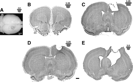
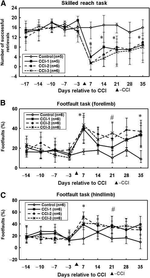
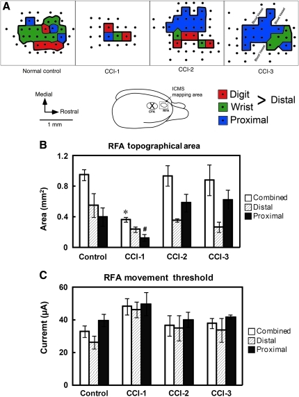
We report the results of controlled cortical impact (CCI) centered on the caudal forelimb area (CFA) of rat motor cortex to determine the feasibility of examining cortical plasticity in a spared cortical motor area (rostral forelimb area, RFA). We compared the effects of three CCI parameter sets (groups CCI-1, CCI-2, and CCI-3) that differed in impactor surface shape, size, and location, on behavioral recovery and RFA structural and functional integrity. Forelimb deficits in the limb contralateral to the injury were evident in all three CCI groups assessed by skilled reach and footfault tasks that persisted throughout the 35-day post-CCI assessment period. Nissl-stained coronal sections revealed that the RFA was structurally intact. Intracortical microstimulation experiments conducted at 7 weeks post-CCI demonstrated that RFA was functionally viable. However, the size of the forelimb representation decreased significantly in CCI-1 compared to the control group. Subdivided into component movement categories, there was a significant group effect for proximal forelimb movements. The RFA area reduction and reorganization are discussed in relation to possible diaschisis, and to compensatory functional behavior, respectively. Also, an inverse correlation between the anterior extent of the lesion and the size of the RFA was identified and is discussed in relation to corticocortical connectivity. The results suggest that CCI can be applied to rat CFA while sparing RFA. This CCI model can contribute to our understanding of neural plasticity in premotor cortex as a substrate for functional motor recovery.


Figures

FIG. 1.
FIG. 1.
(A) Dorsal view of the lesion (group CCI-1). Coronal sections at (B) 2.7 mm from the bregma at the level of the RFA (group CCI-3; representative of all cases); (C) ∼ 0.2 mm from the bregma at the level of the CFA (group CCI-2; representative of majority of cases); (D) ∼ 0.2 mm from the bregma at the level of the CFA (group CCI-3; representative of five cases); (E) ∼0.2 mm from the bregma at the level of the CFA (group CCI-1; representative of five cases). The dorsal view in A corresponds to the injury severity shown in E (scale bar = 1 mm; impactor tip shape [rounded or flat] and size [mm] are indicated by the inset associated with each panel [not to scale]). The rounded tip often resulted in some sparing of deep cortical layers at the boundary of the lesion, while the flat tip typically resulted in a more uniform column of damage. No histological evidence of damage was found at the level of the RFA (B) in any of the cases (CCI, controlled cortical impact; CFA, caudal forelimb area; RFA, rostral forelimb area).
FIG. 2.
FIG. 2.
Histological results. (A) Scatterplot showing distribution of lesion volume in the CFA region in each of the three experimental groups. The three different sets of impact tip parameters produced no significant differences in lesion volume. Means are indicated by horizontal lines. (B) Bar graphs showing rostrocaudal extents (±standard error of the mean) measured from the bregma. The three sets of impact tip parameters produced no difference in rostral lesion extent (upper portion of plot), but a significant difference in caudal extent (lower portion of plot). The caudal extent of group CCI-1 was more anterior than that of groups CCI-2 and CCI-3 (*p < 0.05; CCI, controlled cortical impact; CFA, caudal forelimb area).
FIG. 3.
FIG. 3.
Behavioral results. (A) Median number of successful retrievals (±95% confidence intervals) on testing sessions before and after CCI. Deficits in skilled reach were observed during 4 of 5 post-lesion sessions. On post-lesion day 7, the pellet retrievals of the control and CCI groups corresponded to a success rate of >70% and <20%, respectively. The number of successful retrievals of the CCI groups on post-lesion day 35 corresponded to a success rate of 41–48% (*p < 0.05 for each of the three CCI groups compared to the control group). (B) Median percent forelimb footfaults (±95% confidence intervals) on testing sessions before and after CCI. Increased errors in forelimb locomotion occurred in 3 of 5 post-lesion sessions (*p < 0.05 for each of the three CCI groups compared to the control group; #p < 0.05 for groups CCI-2 and CCI-3 compared to the control group). (C) Median percent hindlimb footfaults (±95% confidence intervals) on testing sessions before and after CCI. Increased hindlimb footfault errors occurred in 2 of 5 post-lesion testing sessions (*p < 0.05 for each of the three CCI groups compared to the control group; #p < 0.05 for groups CCI-2 and CCI-3 compared to the control group; CCI, controlled cortical impact).
FIG. 4.
FIG. 4.
Neurophysiological results. (A) Color-coded maps of movements evoked by ICMS in the RFA 7 weeks after CCI. The inset shows the ICMS mapping area illustrated on a dorsolateral view of the rat brain. Cases illustrated are representative of movement representations in each group. Each map was bordered by face, whisker, or neck movements. Dots reflect the ICMS penetration sites. Note the obviously smaller RFA size in CCI-1 animals. In this figure, distal forelimb movements are subdivided further into digit and wrist movements. Note that no digit movements were evoked from any of the CCI-3 animals. In these cases, distal forelimb movements consisted of wrist movements exclusively. (B) Movement representation areas (±standard error of the mean [SEM]) in the RFA. The combined RFA (distal + proximal representations) was smallest in group CCI-1 (*p < 0.05 for group CCI-1 compared to each of the other groups; #p < 0.05 for group CCI-1 compared to groups CCI-2 and CCI-3). (C) Threshold currents required to evoke movements in the RFA (±SEM). Although there appeared to be an overall tendency for higher currents in group CCI-1, no significant difference was detected among groups (CCI, controlled cortical impact; ICMS, intracortical microstimulation; RFA, rostral forelimb area).
FIG. 5.
FIG. 5.
Neurophysiologically-based RFA area as a function of rostral lesion extent. The more rostral the lesion extended, that is, the closer the lesion was to the RFA, the smaller the RFA area obtained during the post-CCI mapping procedure (diamonds). Three cases in which we could not evoke movements (and therefore were excluded from the statistical analysis) are also illustrated (circle). Numbers indicate CCI groups (CCI, controlled cortical impact; RFA, rostral forelimb area).

References

    1. Adkins D.L. Jones T.A. D-amphetamine enhances skilled reaching after ischemic cortical lesions in rats. Neurosci. Lett. 2005;380:214–218. - PubMed
    1. Alaverdashvili M. Foroud A. Lim D.H. Whishaw I.Q. “Learned baduse” limits recovery of skilled reaching for food after forelimb motor cortex stroke in rats: a new analysis of the effect of gestures on success. Behav. Brain Res. 2008;188:281–290. - PubMed
    1. Alaverdashvili M. Lim D.H. Whishaw I. Q. No improvement by amphetamine on learned non-use, attempts, success or movement in skilled reaching by the rat after motor cortex stroke. Eur. J. Neurosci. 2007;25:3442–3452. - PubMed
    1. Allred R.P. Adkins D.L. Woodlee M.T. Husbands L.C. Maldonado M.A. Kane J.R. Schallert T. Jones T.A. The vermicelli handling test: a simple quantitative measure of dexterous forepaw function in rats. J. Neurosci. Methods. 2008;170:229–244. - PMC - PubMed
    1. Barth T.M. Jones T.A. Schallert T. Functional subdivisions of the rat somatic sensorimotor cortex. Behav. Brain Res. 1990;39:73–95. - PubMed

Publication types