

Resolvin E1 improves tear production and decreases inflammation in a dry eye mouse model
- PMID: 20874497
- PMCID: PMC2956380
- DOI: 10.1089/jop.2010.0019
Resolvin E1 improves tear production and decreases inflammation in a dry eye mouse model
Abstract
Purpose: Dry eye (DE) is a common ocular surface disease, particularly among women and the elderly, with chronic symptoms of eye irritation and, in severe cases, blurred vision. Several studies have shown that there is an inflammatory component in DE, although the pathogenesis is not thoroughly understood. Resolvin E1 (RvE1; RX-10001) is an endogenous mediator derived from the omega-3 polyunsaturated fatty acid eicosapentaenoic acid and is involved in inflammation resolution and tissue protection. Here we investigated the role of RvE1 in a DE mouse model.
Methods: Thirteen- to 14-week-old female BALB/C mice were exposed to desiccating conditions. One week after DE exposure, animals were treated topically with drug or vehicle 4 times per day for an additional week. Controls were nontreated animals placed in a normal environment. Schirmer's test was performed before treatment initiation and at days 2 and 4 after treatment. Density of corneal epithelial cells was analyzed in vivo using the Rostock Cornea Module of the Heidelberg Retina Tomograph (HRT-II). Corneas were processed using Western blot analysis and immunofluorescence examination.
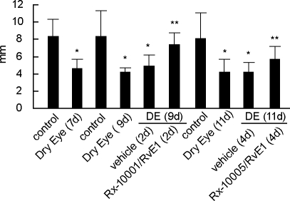
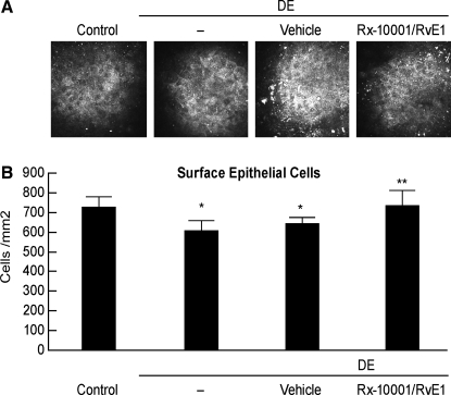
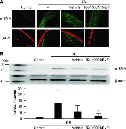
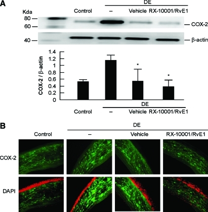
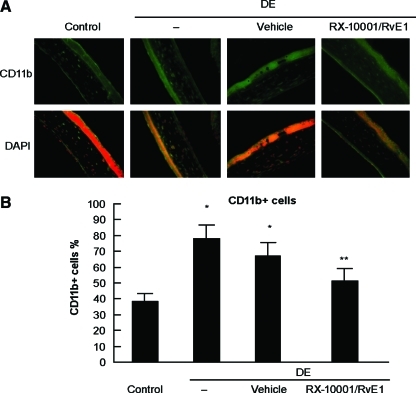
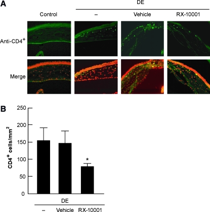
Results: Schirmer's test showed a significant decrease in tear production in DE compared with controls. There was no change at 2 and 4 days after treatment with the vehicle, but a significant increase was observed at 2 and 4 days in the RvE1-treated group. The density of the superficial epithelial cells showed a significant decrease after DE compared with controls, which increased after 7 days of RvE1 treatment. Western blot analysis showed that α-smooth muscle actin and cyclooxygenase-2 (COX-2) expression were strongly upregulated after DE and decreased after 7 days of RvE1 treatment. Immunofluorescence confirmed strong positive staining of α-smooth muscle actin and COX-2 in stroma and/or in epithelia after DE, which decreased with RvE1 treatment. The percentage of infiltrating CD⁴+ T cells and CD11b+ cells decreased after RvE1 treatment when compared with DE.
Conclusion: RvE1 promotes tear production, corneal epithelial integrity, and a decrease in inflammatory inducible COX-2. In the stroma, RvE1 inhibits keratocyte transformation to myofibroblasts and lowers the number of monocytes/macrophages in this DE mouse model. These results suggest that RvE1 and similar resolvin analogs have therapeutic potential in the treatment of DE.
Figures
References
-
- Smith J.A. Albeitz J. Begley C., et al. The epidemiology of dry eye disease: Report of the Epidemiology Subcommittee of the International Dry Eye Workshop. Ocul. Surf. 2007;5:93–107. - PubMed
-
- Schaumberg D.A. Sullivan D.A. Dana M.R. Epidemiology of dry eye syndrome. Adv. Exp. Med. Biol. 2002;506(Pt B):989–998. - PubMed
-
- Stern M.E. Beuerman R.W. Fox R.I., et al. The pathology of dry eye: the interaction between the ocular surface and lacrimal glands. Cornea. 1998;17:584–589. - PubMed
-
- Luo L. Li D.Q. Doshi A., et al. Experimental dry eye stimulates production of inflammatory cytokines and MMP-9 and activates MAPK signaling pathways on the ocular surface. Invest. Ophthalmol. Vis. Sci. 2004;45:4293–4301. - PubMed
-
- Perry H.D. Solomon R. Donnenfeld E.D., et al. Evaluation of topical cyclosporine for the treatment of dry eye disease. Arch. Ophthalmol. 2008;126:1046–1050. - PubMed
Publication types
MeSH terms
Substances
Grants and funding
LinkOut - more resources
Full Text Sources
Other Literature Sources
Research Materials
