. 2010 Dec;163(6):1218-28.
doi: 10.1111/j.1365-2133.2010.10063.x.

In vivo reflectance confocal microscopy of shave biopsy wounds: feasibility of intraoperative mapping of cancer margins

Affiliations

In vivo reflectance confocal microscopy of shave biopsy wounds: feasibility of intraoperative mapping of cancer margins

A Scope et al. Br J Dermatol. 2010 Dec.

Abstract

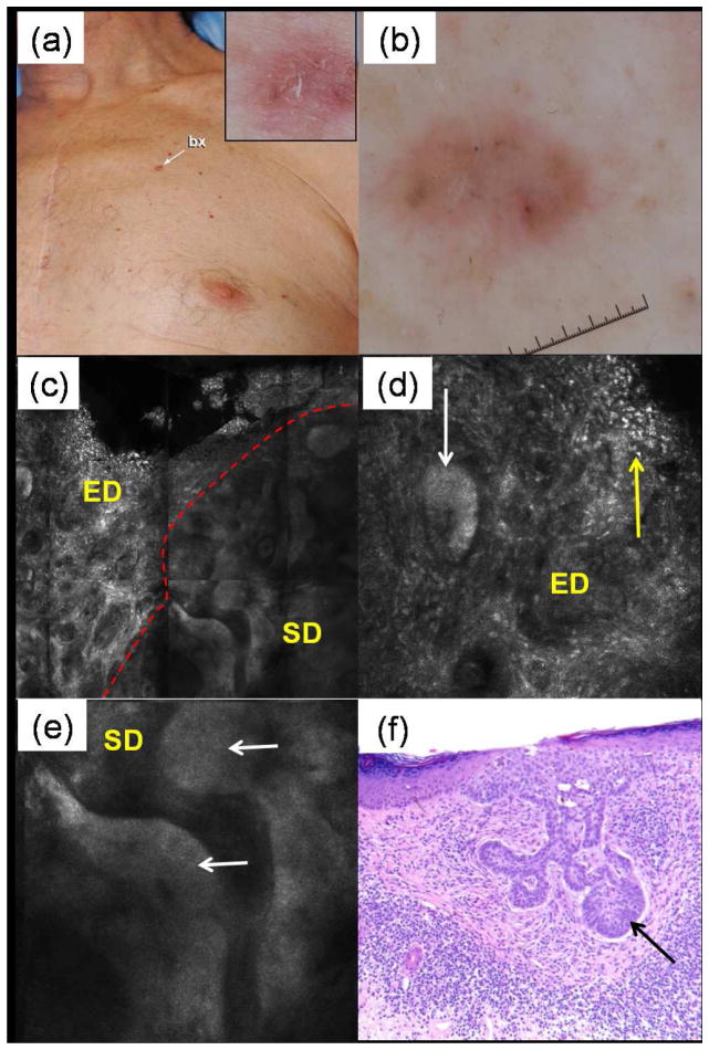
Background: Reflectance confocal microscopy (RCM) images skin at cellular resolution and has shown utility for the diagnosis of nonmelanoma skin cancer in vivo. Topical application of aluminium chloride (AlCl(3)) enhances contrast in RCM images by brightening nuclei.

Objectives: To investigate feasibility of RCM imaging of shave biopsy wounds using AlCl(3) as a contrast agent.

Methods: AlCl(3) staining was optimized, in terms of concentration vs. immersion time, on excised tissue ex vivo. RCM imaging protocol was tested in patients undergoing shave biopsies. The RCM images were retrospectively analysed and compared with the corresponding histopathology.

Results: For 35% AlCl(3) , routinely used for haemostasis in clinic, minimum immersion time was determined to be 1 min. We identified three consistent patterns of margins on RCM mosaic images by varying depth: epidermal margins, peripheral dermal margins, and deep dermal margins. Tumour islands of basal cell carcinoma were identified at peripheral or deep dermal margins, correlating on histopathology with aggregates of neoplastic basaloid cells. Atypical cobblestone or honeycomb patterns were identified at the epidermal margins in squamous cell carcinomas, correlating with a proliferation of atypical keratinocytes extending to biopsy margins.

Conclusions: RCM imaging of shave biopsy wounds is feasible and demonstrates the future possibility of intraoperative mapping in surgical wounds.


Conflict of interest statement

Conflicts of interest: Dr. Milind Rajadhyaksha is a former employee and owns equity in Lucid Inc., the company that makes and sells the VivaScope confocal microscope. The VivaScope is the commercial version of an original laboratory prototype of a confocal scanning laser microscope that was developed by Dr. Rajadhyaksha when he was in the Department of Dermatology at Massachusetts General Hospital, Harvard Medical School. All other authors have no conflicts of interest to declare.

Figures

Figure 1
Figure 1
Setup for attachment of reflectance confocal microscope (RCM) objective lens to shave biopsy wound. The skin at the site of imaging includes the surgical wound cavity with adjacent intact surrounding epidermis (“se”) and dermis (“sd”). The surgical wound cavity is filled with sterile gel and covered by transparent sterile dressing. Following the application of an oil drop onto the dressing, the tissue ring is attached to the dressing, to cover both a portion of the wound cavity and its margins. The top, concave side of the tissue ring is filled with ultrasound (“US”) gel as the immersion medium. The tissue ring serves as a docking template for the RCM objective lens for locating and stabilizing the site to be imaged. The three depth levels of en face RCM imaging are shown, including the “epidermal margin” at the level of the surrounding epidermis, “peripheral dermal margin” at the level of the surrounding superficial dermis, and “deep dermal margin” at the level of the base of the wound.
Figure 2
Figure 2
Aluminum chloride (AlCl3)-stained epidermis showing nuclear brightening and enhanced nuclei-to-dermis contrast for different immersion times and AlCl3 concentrations. The epidermis is vertically-sectioned. (a) Unstained control; (b) 20% AlCl3, 3 minutes of immersion; (c) 35% AlCl3, 1 minute of immersion; and (d) 50% AlCl3 in water, 10 seconds of immersion. While the unstained image (a) shows a honeycomb pattern of dark nuclei and bright cellular outlines in the epidermis as normally seen with in vivo RCM, all AlCl3-stained tissue specimens (b–d) present a cobblestone pattern of bright nuclei in the epidermis.
Figure 3
Figure 3
Histopathological correlation of RCM features seen at shave biopsy margins. (a) Nuclei of keratinocytes that line the wound cavity appear bright, following application of aluminum chloride; this is referred to as cobblestone pattern. (b) Normal keratinocytes in the surrounding epidermis adjacent to the wound cavity display a different pattern, referred to as honeycomb pattern, whereby the nuclei are dark and the cellular outlines of keratinocytes are bright; (c) both patterns correlate with spinous and granular layers without atypia of keratinocytes. (d) Honeycomb pattern adjacent to cobblestone pattern; the dashed line of demarcation indicates the edge of the wound at the level of the epidermal margin. (e) Bright linear and curved structures in the dermis with a background of amorphous brightness seen on RCM correlated with collagen bundles in solar-altered dermis on histopathology (f). (g) Small round bright features and bright stellate cells in the dermis seen on RCM correlated with an infiltrate of lymphocytes and histiocytes on histopathology (h). Scale = 100μm on RCM images.
Figure 4
Figure 4
Wound margins at varying depths. The level of imaging is depicted by the dashed line in the drawings at the top of each column. (a–b) Epidermal margin; the RCM mosaic (a, 4×4 mm) is taken at the level of the spinous layer of the epidermis. The surrounding epidermis (“SE”) displays a honeycomb pattern. The dark area represents the gel-filled wound cavity. At higher magnification RCM (b, 0.5×0.5 mm) a regular honeycomb pattern in the surrounding epidermis (yellow arrow) can be seen, as well as few bright nuclei in cobblestone pattern within the wound cavity (white arrow). The demarcation between honeycomb and cobblestone patterns, denoting the wound edge, is shown (yellow dashed line). (c–d) Peripheral dermal margin; in this RCM mosaic (c, 4×4 mm) the surrounding skin (‘SS”) is blurred since imaging is performed through intact stratum corneum, resulting in loss of backscattered detected light with increasing depth. In contrast, the exposed dermis (“ED”) in the wound shows bright reticulated collagen of the papillary dermis. The base of the wound is still below the plane of imaging and therefore appears dark. At higher magnification RCM (d, 0.5×0.5 mm), there is demarcation (dashed yellow line) between the blurred surrounding skin (asterisk) and the exposed wound tissue showing bright dermal-epidermal junction (white dashed arrows) as well as bright, in-focus collagen in the superficial dermis (dashed yellow arrows). (e–f) Deep dermal margin; on RCM mosaic (e, 4×4 mm), the surrounding dermis appears blurred and dark because of deeper imaging level. However, the RCM focal plane reaches the exposed dermis (“ED”) at the base of the wound, which appears bright. At higher magnification RCM (f, 0.5×0.5 mm) the bright collagen bundles at the base of the wound can be seen (dashed yellow arrow).
Figure 5
Figure 5
Basal cell carcinoma (BCC). Images a&b are from one lesion, images c&d from another lesion. (a) RCM imaging at the level of the dermal-epidermal junction (peripheral dermal margin) shows an aggregate of neoplastic cells appearing as a focus of bright nuclei (asterisk), well delineated from the adjacent epidermis (“E”) and dermis (“D”). (b) An aggregate of basaloid cells with peripheral palisading of nuclei (asterisk) is emanating from the undersurface of the epidermis (“E”) and protruding into the superficial dermis (“D”). The diagnosis is superficial BCC. (c) RCM imaging at the level of the dermis (deep dermal margin) shows a dermal aggregate of neoplastic cells (‘T”) displaying focal peripheral palisading of nuclei (solid arrow); the aggregates are well demarcated from the surrounding dermis by dark clefts (dashed arrow). (d) On histopathology, aggregate of basaloid cells (“T”) are seen in the reticular dermis with palisading of nuclei (solid arrow) and subtle clefting (dashed arrow), diagnostic of nodular BCC. Notably, the tumours were transected during biopsy and appear at the exposed wound surface. Since the neoplastic epithelial cells have been exposed to aluminum chloride application, the nuclei appear bright and help to delineate the tumor aggregates in RCM images.
Figure 6
Figure 6
Basal cell carcinoma (BCC). (a) This patient presented an 8 mm papule on the chest (inset shows close-up clinical image); (b) dermoscopy revealed gray dots and gray-brown ovoid nests, suspicious for BCC. (c) RCM mosaic (1.5×1.5 mm) of the shave biopsy wound is shown at the peripheral dermal margin level; the red dashed line demarcates the darker surrounding dermis (“SD”) from the brighter exposed dermis (“ED”). (d) Individual RCM image (500×500 μm) at the same level showing a bright tumor aggregate (white arrow) in the exposed dermis (“ED”) that also displays numerous bright spots and bright stellate cells (yellow arrow). (e) Individual RCM image (500×500 μm) in the surrounding dermis (“SD”) at the same level also showing tumor aggregates (white arrows). Note that these tumor aggregates and the surrounding dermis (e) appear less bright and with lower resolution than in the exposed dermis (d). (f) On histopathology, an aggregate of basaloid cells with peripheral palisading of nuclei (arrow) is emanating from the undersurface of the epidermis, diagnostic of superficial BCC. Note the dense inflammatory infiltrate which correlates with the numerous bright spots and bright stellate cells seen on RCM.
Figure 7
Figure 7
Squamous cell carcinoma (SCC). (a) This patient presented with a 1.5 cm red, keratotic plaque on the back. (b) A close-up clinical image post shave biopsy showing in the remaining scale in the surrounding skin (arrow). (c) Dermoscopy showing a pink blush and dotted vessels (arrow) in the skin surrounding the shave biopsy wound. (d) RCM mosaic (1.5×1.5 mm) at the level of epidermal margin (level indicated in the drawing above) showing a honeycomb pattern in the surrounding epidermis (“SE”), to the left of the dashed line. Note the enhanced brightness of nuclei in the exposed epidermis (“EE”, right of dashed line) producing a cobblestone pattern. (e) RCM mosaic (1.5×1.5 mm) at the level of peripheral dermal margin (level indicated in the drawing above). Both surrounding dermis (“SD”) and surrounding epidermis (“SE”) are seen to the left of the dashed line, indicating imaging is at the level of the dermal-epidermal junction. In the wound cavity, a cobblestone pattern is seen (asterisk) as well as dermal papillae (arrow). (f) RCM mosaic (1.5×1.5 mm) at the level of deep dermal margin, base of the wound (level indicated in the drawing above). The finding of cobblestone pattern (asterisk) in the dermis where blood vessels can be seen (dashed arrow), is clearly abnormal. (g) RCM individual image (500×500 μm), akin to higher magnification microscopy, of the surrounding skin at epidermal margin level shows an atypical honeycomb pattern, with outlines that vary in thickness and brightness and dark holes (dashed red arrow) that vary in size and shape. There are cells that appear wholly bright, without central dark nucleus (solid red arrow). (h) RCM individual image (500×500 μm), of the exposed epidermis at the epidermal margin level shows an atypical cobblestone pattern, with bright nuclei of keratinocytes (yellow arrows) that are irregularly crowded and display variability in the size of nuclei. (i) On histopathology, there is full thickness atypia of keratinocytes with jumbling, crowding and pleomorphism of nuclei, hyperchromatic nuclei and necrotic keratinocytes (black arrow), diagnostic of SCC. The proliferation of atypical keratinocytes extended to the peripheral margins (“PM”) and base of the biopsy (“base”).

Similar articles

Cited by

References

    1. Bialy TL, Whalen J, Veledar E, et al. Mohs micrographic surgery vs traditional surgical excision: a cost comparison analysis. Arch Dermatol. 2004;140:736–42. - PubMed
    1. Rogers HW, Weinstock MA, Harris AR, et al. Incidence estimate of nonmelanoma skin cancer in the United States, 2006. Arch Dermatol. 2010;146:283–7. - PubMed
    1. Rajadhyaksha M, Grossman M, Esterowitz D, et al. In vivo confocal scanning laser microscopy of human skin: melanin provides strong contrast. J Invest Dermatol. 1995;104:946–52. - PubMed
    1. Rajadhyaksha M, González S, Zavislan JM, et al. In vivo confocal scanning laser microscopy of human skin II: advances in instrumentation and comparison with histology. J Invest Dermatol. 1999;113:293–303. - PubMed
    1. Nori S, Rius-Díaz F, Cuevas J, et al. Sensitivity and specificity of reflectance-mode confocal microscopy for in vivo diagnosis of basal cell carcinoma: a multicenter study. J Am Acad Dermatol. 2004;51:923–30. - PubMed

Publication types