. 2010 Dec;217(6):665-78.
doi: 10.1111/j.1469-7580.2010.01303.x. Epub 2010 Sep 28.

Germ layer differentiation during early hindgut and cloaca formation in rabbit and pig embryos

Affiliations

Germ layer differentiation during early hindgut and cloaca formation in rabbit and pig embryos

Romia Hassoun et al. J Anat. 2010 Dec.

Abstract

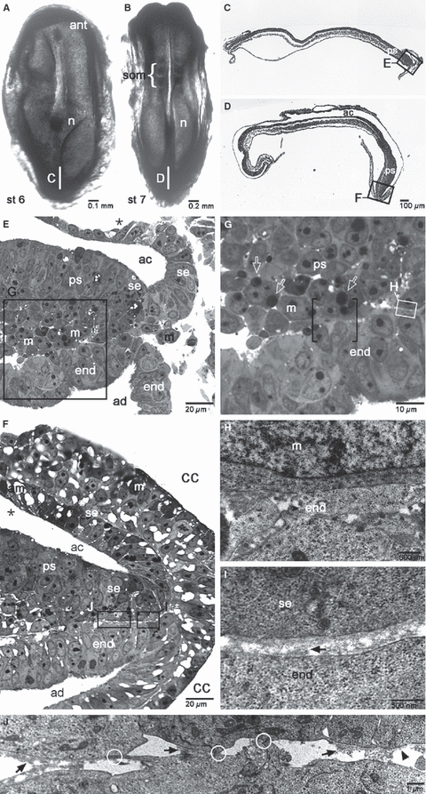
Relative to recent advances in understanding molecular requirements for endoderm differentiation, the dynamics of germ layer morphology and the topographical distribution of molecular factors involved in endoderm formation at the caudal pole of the embryonic disc are still poorly defined. To discover common principles of mammalian germ layer development, pig and rabbit embryos at late gastrulation and early neurulation stages were analysed as species with a human-like embryonic disc morphology, using correlative light and electron microscopy. Close intercellular contact but no direct structural evidence of endoderm formation such as mesenchymal-epithelial transition between posterior primitive streak mesoderm and the emerging posterior endoderm were found. However, a two-step process closely related to posterior germ layer differentiation emerged for the formation of the cloacal membrane: (i) a continuous mesoderm layer and numerous patches of electron-dense flocculent extracellular matrix mark the prospective region of cloacal membrane formation; and (ii) mesoderm cells and all extracellular matrix including the basement membrane are lost locally and close intercellular contact between the endoderm and ectoderm is established. The latter process involves single cells at first and then gradually spreads to form a longitudinally oriented seam-like cloacal membrane. These gradual changes were found from gastrulation to early somite stages in the pig, whereas they were found from early somite to mid-somite stages in the rabbit; in both species cloacal membrane formation is complete prior to secondary neurulation. The results highlight the structural requirements for endoderm formation during development of the hindgut and suggest new mechanisms for the pathogenesis of common urogenital and anorectal malformations.


Figures

Fig. 1
Fig. 1
Light and electron microscopy of germ layer differentiation in the pig at stage 4 as seen in dorsal views of the whole embryonic disc (A), in sagittal semithin (1 μm) (B–D) and ultrathin (E) sections taken from the embryo shown in A at the position marked with a vertical white bar. The position of the areas shown at high magnification is indicated with boxes in pictures of the next lower magnification. Brackets mark the patches of isoprismatic hypoblast at stage 4+, asterisks mark the embryonic-extra-embryonic border in the dorsal layer of the embryonic disc, arrowheads point to the basement membrane, arrows point to patches of flocculent electron-dense material in the extracellular space next to the basement membrane. ant, anterior pole; b, bottle cells of the primitive streak; exm, extra-embryonic mesoderm, m, mesoderm cells; n, node; pm, prechordal mesoderm; ps, primitive streak; se, surface epithelium; tb, trophoblast; vl, ventral layer; yse, yolk sac epithelium.
Fig. 2
Fig. 2
Light and electron microscopy of germ layer differentiation in the pig at stages 6 and 7 as seen in dorsal views of whole embryonic discs (A, B), in sagittal semithin (1 μm) (C–G) and ultrathin (H–J) sections taken from the embryos shown in A (stage 6) and B (stage 7), respectively, at the positions marked with vertical white bars. The position of the areas shown at high magnification is indicated with boxes in the next lower magnification shown. Brackets mark the close apposition between ectoderm and ventral layer cells, open arrows point to apoptotic bodies in mesoderm cells (in G), white circles mark areas of direct intercellular contact. ac, amniotic cavity; ad, allantoic diverticulum; am, amnion epithelium; cc, chorionic cavity; end, endoderm; exm, extra-embryonic mesoderm; all other labeling as in Fig. 1.
Fig. 3
Fig. 3
Light and electron microscopy of germ layer differentiation in the rabbit at stages 5 and 7 as seen in dorsal views of whole embryonic discs (A,B), in sagittal semithin (1 μm) (C,D) and ultrathin (E,F) sections taken from the same embryos shown in A (stage 5) and B (stage 7), respectively, at the position marked with vertical white bars. The position of areas selected for high magnification is indicated with boxes in the lower magnification shown. Curved brackets in C mark the prospective region of cloacal membrane formation, # marks artefactual fold in the extra-embryonic region in A. All other labelling as in Figs 1 and 2.
Fig. 4
Fig. 4
Light and electron microscopy of germ layer differentiation in the rabbit at stages 8 and 9 as seen in dorsal views of whole embryonic discs (insets in A–C), in transverse (A) and median sagittal (B–D) semithin (1 μm) and ultrathin (E–H) sections taken from the same or similar embryos shown in insets. The position of the section from the embryo shown in A is marked in the inset with a short horizontal white line, the position of the areas selected for high magnification is indicated with boxes in the lower magnification shown. i, invagination of ventral layer cells, cap, capillary; all other labelling as in Figs 1, 2 and 3. Magnification bars in insets: 500 μm.
Fig. 5
Fig. 5
Schematic median sagittal sections showing germ layer differentiation and fate at the posterior pole of the mammalian embryo from late blastocyst stages (A), through stages 1 and 2 of cloacal membrane formation (gastrulation and primary neurulation, B and C, respectively) until the start of hindgut closure and tail bud formation (and secondary neurulation) during early organogenesis (D). The position of the sections is indicated in the embryonic disc outlines of appropriate stages in A, B and C. The colour scheme illustrates the equivalence structures and their fate in subsequent stages, i.e. the dark blue of the epiblast overlying the primitive streak at stages 1 and 2 of cloacal membrane formation (B and C) has disappeared during tail bud formation and hindgut closure (in D) because the epiblast is thought to generate surface ectoderm, neuroectoderm (dark blue, now with – olive-shaded – lumen), mesoderm and endoderm of the tail. The broken arrows in C and D indicate the longitudinal axis of the emerging and extending tail bud, respectively. The stages shown in B and C were investigated in this study; A and D are shown to illustrate the topography of the origin and the fate of the germ layers involved, respectively. D shows the situation after further ventral bending of the posterior margin of the embryonic disc and after emergence of the tail bud from the dorsal aspect of the primitive streak. Through both these processes, the allantoic diverticulum (and with it the connecting stalk mesoderm and the amniotic covering of the umbilical cord) is carried ventrally and the cloacal membrane rotates by about 130° into an almost frontal position. For the sake of simplicity the amniotic membrane, with its roots at the umbilical cord and not covering the tail bud, is shown in D only. To avoid confusion, the thickness of germ layers is not drawn to scale.

Similar articles

Cited by

References

    1. Blum M, Andre P, Muders K, et al. Ciliation and gene expression distinguish between node and posterior notochord in the mammalian embryo. Differentiation. 2007;75:133–146. - PubMed
    1. Burtscher I, Lickert H. Foxa2 regulates polarity and epithelialization in the endoderm germ layer of the mouse embryo. Development. 2009;136:1029–1038. - PubMed
    1. Charrier J, Teillet M, Lapointe F, et al. Defining subregions of Hensen's node essential for caudalward movement, midline development and cell survival. Development. 1999;126:4771–4783. - PubMed
    1. Collombat P, Xu X, Ravassard P, et al. The ectopic expression of Pax4 in the mouse pancreas converts progenitor cells into alpha and subsequently beta cells. Cell. 2009;138:449–462. - PMC - PubMed
    1. Downs KM. Systematic localization of oct-3/4 to the gastrulating mouse conceptus suggests manifold roles in mammalian development. Dev Dyn. 2008;237:464–475. - PubMed

Publication types

LinkOut - more resources