

Platelet autologous growth factors decrease the osteochondral regeneration capability of a collagen-hydroxyapatite scaffold in a sheep model
- PMID: 20875101
- PMCID: PMC2954989
- DOI: 10.1186/1471-2474-11-220
Platelet autologous growth factors decrease the osteochondral regeneration capability of a collagen-hydroxyapatite scaffold in a sheep model
Abstract
Background: Current research aims to develop innovative approaches to improve chondral and osteochondral regeneration. The objective of this study was to investigate the regenerative potential of platelet-rich plasma (PRP) to enhance the repair process of a collagen-hydroxyapatite scaffold in osteochondral defects in a sheep model.
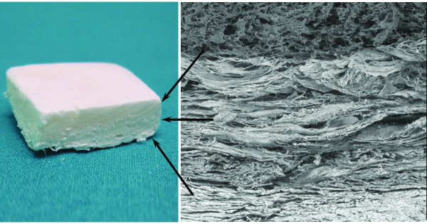
Methods: PRP was added to a new, multi-layer gradient, nanocomposite scaffold that was obtained by nucleating collagen fibrils with hydroxyapatite nanoparticles. Twenty-four osteochondral lesions were created in sheep femoral condyles. The animals were randomised to three treatment groups: scaffold, scaffold loaded with autologous PRP, and empty defect (control). The animals were sacrificed and evaluated six months after surgery.
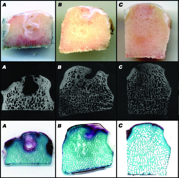
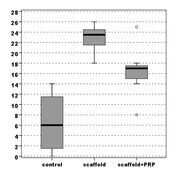
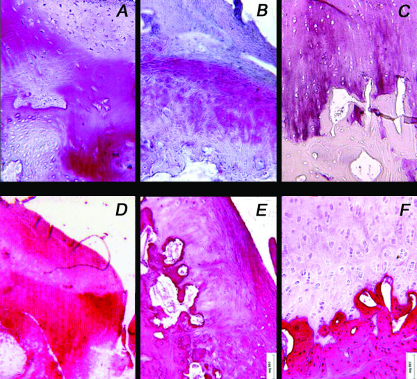
Results: Gross evaluation and histology of the specimens showed good integration of the chondral surface in both treatment groups. Significantly better bone regeneration and cartilage surface reconstruction were observed in the group treated with the scaffold alone. Incomplete bone regeneration and irregular cartilage surface integration were observed in the group treated with the scaffold where PRP was added. In the control group, no bone and cartilage defect healing occurred; defects were filled with fibrous tissue. Quantitative macroscopic and histological score evaluations confirmed the qualitative trends observed.
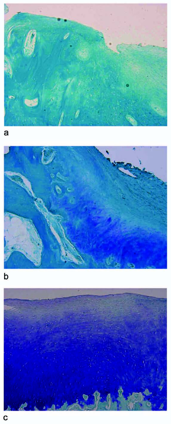
Conclusions: The hydroxyapatite-collagen scaffold enhanced osteochondral lesion repair, but the combination with platelet growth factors did not have an additive effect; on the contrary, PRP administration had a negative effect on the results obtained by disturbing the regenerative process. In the scaffold + PRP group, highly amorphous cartilaginous repair tissue and poorly spatially organised underlying bone tissue were found.
Figures


References
-
- Bartlett W, Skinner JA, Gooding CR, Carrington RWJ, Flanagan AM, Briggs TWR, Bentley G. Autologous chondrocyte implantation versus matrix-induced autologous chondrocyte implantation for osteochondral defects of the knee: a prospective, randomised study. J Bone Joint Surg Br. 2005;87:640–5. doi: 10.1302/0301-620X.87B5.15905. - DOI - PubMed
-
- Schumann D, Ekaputra AK, Lam CXF, Hutmacher DW. Biomaterials/scaffolds. Design of bioactive, multiphasic PCL/collagen type I and type II-PCL-TCP/collagen composite scaffolds for functional tissue engineering of osteochondral repair tissue by using electrospinning and FDM techniques. Methods Mol Med. 2007;140:101–24. full_text. - PubMed
-
- Mano JF, Silva GA, Azevedo HS, Malafaya PB, Sousa RA, Silva SS, Boesel LF, Oliveira JM, Santos TC, Marques AP, Neves NM, Reis RL. Natural origin biodegradable systems in tissue engineering and regenerative medicine: present status and some moving trends. J R Soc Interface. 2007;4:999–1030. doi: 10.1098/rsif.2007.0220. - DOI - PMC - PubMed
Publication types
MeSH terms
Substances
LinkOut - more resources
Full Text Sources
Other Literature Sources
Research Materials
