Review
. 2010 Dec;16(6):653-60.
doi: 10.1017/S1431927610093864. Epub 2010 Sep 27.

Contributions of electron microscopy to understand secretion of immune mediators by human eosinophils

Affiliations
Review

Contributions of electron microscopy to understand secretion of immune mediators by human eosinophils

Rossana C N Melo et al. Microsc Microanal. 2010 Dec.

Abstract

Mechanisms governing secretion of proteins underlie the biologic activities and functions of human eosinophils, leukocytes of the innate immune system, involved in allergic, inflammatory, and immunoregulatory responses. In response to varied stimuli, eosinophils are recruited from the circulation into inflammatory foci, where they modulate immune responses through the release of granule-derived products. Transmission electron microscopy (TEM) is the only technique that can clearly identify and distinguish between different modes of cell secretion. In this review, we highlight the advances in understanding mechanisms of eosinophil secretion, based on TEM findings, that have been made over the past years and that have provided unprecedented insights into the functional capabilities of these cells.


Figures
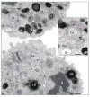
Figure 1
Figure 1. (Color online) Ultrastructure of activated human eosinophils
A: After stimulation with an eosinophil agonist (eotaxin), secretory granules (Gr) exhibit different degrees of emptying of their contents and show morphological diversity indicative of piecemeal degranulation. These granules show disassembled matrices and cores or residual cores or lucent areas in their cores, matrices, or both. Emptying granules are generally larger than resting granules (*), which show typical morphology with a welldefined electron-dense crystalline core. The boxed area shows many profiles of small and large vesicles (EoSVs, highlighted in pink in Ai) in contact and surrounding mobilized secretory granules. Lipid bodies (LB), inflammation-related organelles (Melo et al., 2006), are also formed in response to eotaxin stimulation. Cells were incubated with eotaxin for 1 h, immediately fixed and prepared for TEM as before (Melo et al., 2005a). N, nucleus. Bar: (A) 1.0 μm; (Ai) 600 nm.
Figure 2
Figure 2. (Color online) Secretory granules exhibit internal membranous domains in human eosinophils
A: High magnification of an emptying granule (Gr) with internal membranous tubules (highlighted in blue in Ai) seen in conjunction with mobilized core. EoSVs around the granule are indicated by arrows. A budding vesicle is indicated (arrowhead). Cells were stimulated with eotaxin as before (Melo et al., 2005a) and processed for TEM. Bar, 500 nm. Reprinted from Melo et al. (2005a) with permission.
Figure 3
Figure 3. Secretory granules are highly labeled for CD63 and show membranes organized as a tubular network
A: Immunonanogold electron microscopy revealed pools of CD63 within specific granules undergoing depletion of their contents. The boxed areas show different profiles of tubular vesicles (EoSVs highlighted in pink in higher magnification in Ai and Aii) exhibiting membrane-associated labeling for CD63. Labeling for this tetraspanin is also observed at cell surface (arrows). B, C: Show 3D models generated from serial tomographic slices (~4 nm thick) obtained from a stimulated eosinophil secretory granule analyzed by automated electron tomography (Melo et al., 2005a). The outer granule membrane is partially traced in red and intragranular vesiculotubular membranes in blue. Bar: (A) 600 nm; (Ai, Aii) 300 nm; (B) 400 nm; (C) 150 nm. Panels B and C were reprinted from Melo et al. (2005a) with permission.
Figure 4
Figure 4. EoSVs are formed from mobilized secretory granules in activated eosinophils
A–C: Representative serial tomographic slices (4 nm thick) obtained from electron tomography of a mobilized granule show substantial changes associated with EoSV formation. EoSVs, the lumens of which are highlighted in pink in panels Ai–Ci are imaged as open, tubular-shaped structures. Numbers on the lower right corners of the panels indicate slice numbers through the tomographic volume. D, E: 3D models, based on electron tomographic slices, show the structural organization of EoSVs (colored in pink). D: The granule limiting membrane is highlighted in blue and intragranular membranes in green. E: The model shows the corresponding boxed area in panel Ci. F, G: Immunonanogold EM for major basic protein (F) and interleukin-4 (G) reveals labeling at EoSV lumen and membranes, respectively. Note that the MBP-positive EoSV is fused with the plasma membrane (arrow) in panel F. Cells were stimulated with eotaxin as described (Melo et al., 2005b). Gr, secretory granule. Bars: (A–C) 500 nm; (D) 800 nm; (E) 150 nm; (F, G) 200 nm. Figures 4A–C,E were reprinted from Melo et al. (2005b), Figure 4F from Melo et al. (2009), and Figure 4G from Spencer et al. (2006) with permission.

Similar articles

Cited by

References

    1. Ackerman SJ, Bochner BS. Mechanisms of eosinophilia in the pathogenesis of hypereosinophilic disorders. Immunol Allergy Clin North Am. 2007;27(3):357–375. - PMC - PubMed
    1. Adamko DJ, Odemuyiwa SO, Vethanayagam D, Moqbel R. The rise of the phoenix: The expanding role of the eosinophil in health and disease. Allergy. 2005;60(1):13–22. - PubMed
    1. Ahlstrom-Emanuelsson CA, Greiff L, Andersson M, Persson CG, Erjefalt JS. Eosinophil degranulation status in allergic rhinitis: Observations before and during seasonal allergen exposure. Eur Respir J. 2004;24(5):750–757. - PubMed
    1. Akuthota P, Wang HB, Spencer LA, Weller PF. Immunoregulatory roles of eosinophils: A new look at a familiar cell. Clin Exp Allergy. 2008;38(8):1254–1263. - PMC - PubMed
    1. Armengot M, Garin L, Carda C. Eosinophil degranulation patterns in nasal polyposis: An ultrastructural study. Am J Rhinol Allergy. 2009;23(5):466–470. - PubMed

Publication types

MeSH terms