

KSR1 protects from interleukin-10 deficiency-induced colitis in mice by suppressing T-lymphocyte interferon-γ production
- PMID: 20875416
- PMCID: PMC3008308
- DOI: 10.1053/j.gastro.2010.09.041
KSR1 protects from interleukin-10 deficiency-induced colitis in mice by suppressing T-lymphocyte interferon-γ production
Abstract
Background & aims: Immunological disorders of the gastrointestinal tract such as inflammatory bowel disease often result in recurrent and persistently elevated levels of proinflammatory cytokines. Kinase suppressor of Ras 1 (KSR1) is involved in tumor necrosis factor-mediated colon epithelial cell survival, yet its role in chronic inflammation has not been defined. In this study, we tested the hypothesis that KSR1 is protective against spontaneous experimental colitis.
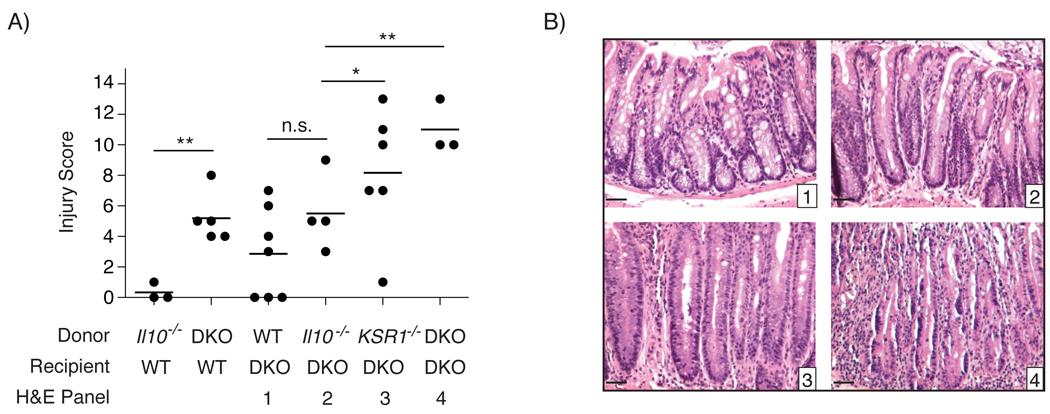
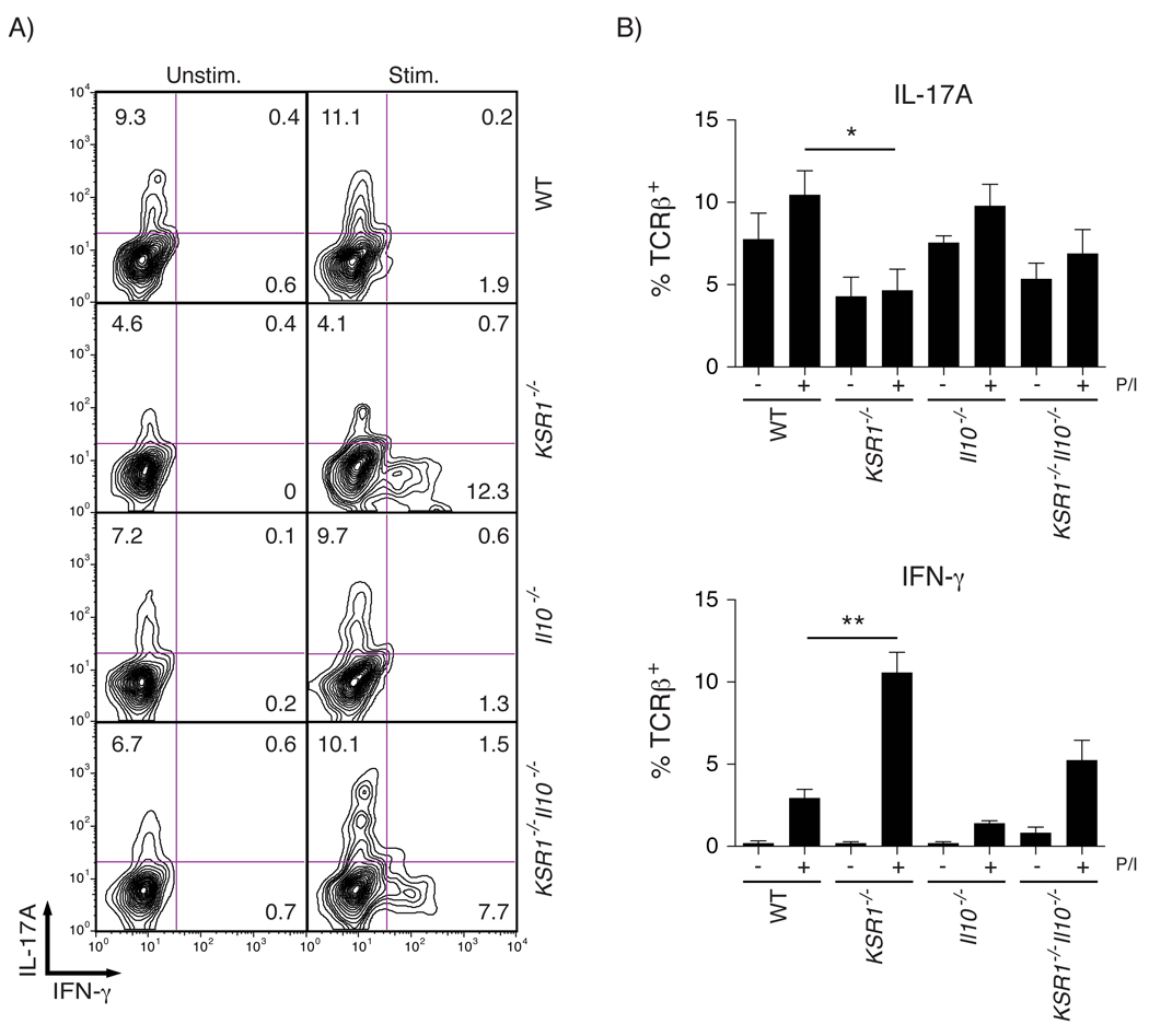
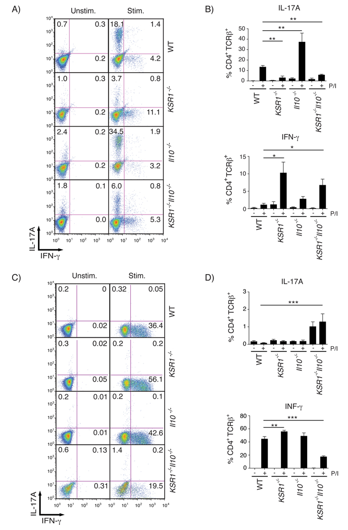
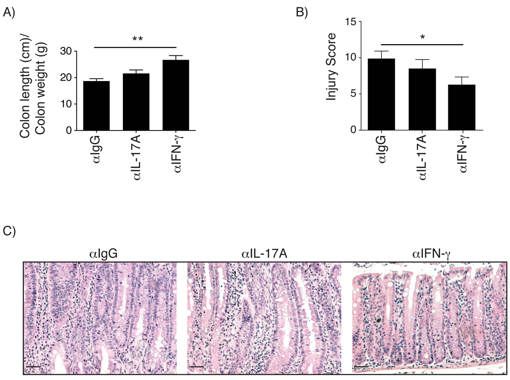
Methods: KSR1(-/-)Interleukin-10 (Il10)(-/-) mice were generated and histolopathologic parameters of intestinal inflammation were scored. Bone marrow transplants performed on wild-type and KSR1(-/-)Il10(-/-) mice determined the contribution of KSR1 in hematopoietic lineages. Mucosal T helper (Th) 1 and Th17 cytokine were also examined. In vitro Th1 and Th17 polarization assays were conducted and interleukin (IL)-17A and interferon-γ (IFN-γ) production analyzed by flow cytometry. Neutralizing antibodies against IgG, IL-17A, or IFN-γ were administered to 3-week-old KSR1(-/-)Il10(-/-) mice for 3 weeks and scored for colitis.
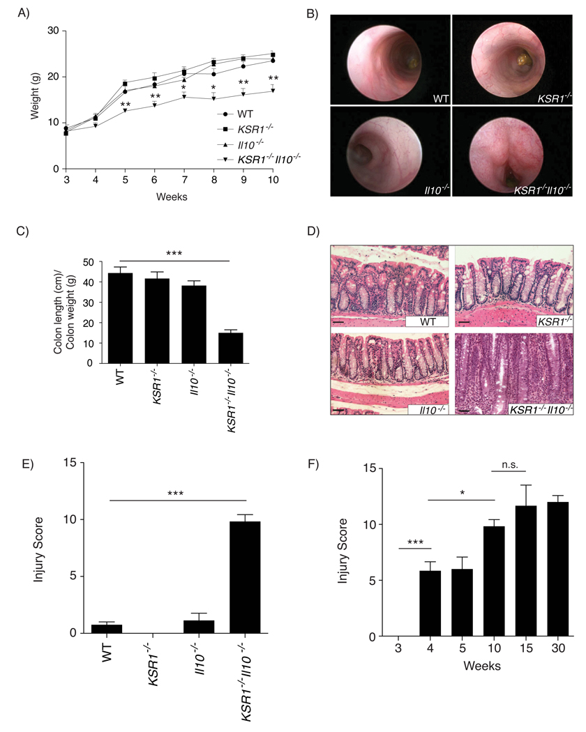
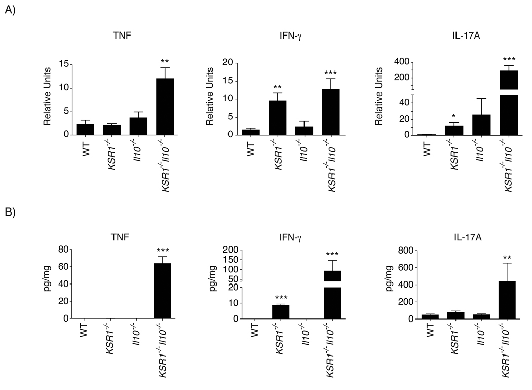
Results: KSR1(-/-)Il10(-/-) mice developed accelerated and severe spontaneous colitis by 4 weeks of age. KSR1 expression in hematopoietic lineages was protective against colitis. Both IFN-γ and IL-17A transcripts were elevated in colons of KSR1(-/-) and KSR1(-/-)Il10(-/-) mice. IFN-γ production was increased in lamina propria T cells isolated from KSR1(-/-) and KSR1(-/-)Il10(-/-) mice. Additionally, in vitro Th1 polarization was increased while Th17 polarization was impaired in KSR1-deficient naïve T cells. Finally, administration of IFN-γ neutralizing antibodies attenuated colitis in KSR1(-/-)Il10(-/-) mice.
Conclusions: Mice lacking both KSR1 and IL-10 develop exacerbated colitis due to dysregulated IFN-γ production in T lymphocytes.
Copyright © 2011. Published by Elsevier Inc.
Conflict of interest statement
Figures
Similar articles
-
Colitis and intestinal inflammation in IL10-/- mice results from IL-13Rα2-mediated attenuation of IL-13 activity.Gastroenterology. 2011 Jan;140(1):254-64. doi: 10.1053/j.gastro.2010.09.047. Epub 2010 Oct 14. Gastroenterology. 2011. PMID: 20951137 Free PMC article.
-
Reciprocal regulation of the survival and apoptosis of Th17 and Th1 cells in the colon.Inflamm Bowel Dis. 2012 Feb;18(2):333-43. doi: 10.1002/ibd.21772. Epub 2011 May 25. Inflamm Bowel Dis. 2012. PMID: 21618360
-
Th17 cells induce colitis and promote Th1 cell responses through IL-17 induction of innate IL-12 and IL-23 production.J Immunol. 2011 Jun 1;186(11):6313-8. doi: 10.4049/jimmunol.1001454. Epub 2011 Apr 29. J Immunol. 2011. PMID: 21531892 Free PMC article.
-
Reciprocal IFN-gamma and TGF-beta responses regulate the occurrence of mucosal inflammation.Immunol Today. 1997 Feb;18(2):61-4. doi: 10.1016/s0167-5699(97)01000-1. Immunol Today. 1997. PMID: 9057354 Review.
-
The Th1 life cycle: molecular control of IFN-γ to IL-10 switching.Trends Immunol. 2011 Jun;32(6):278-86. doi: 10.1016/j.it.2011.03.010. Epub 2011 Apr 30. Trends Immunol. 2011. PMID: 21531623 Review.
Cited by
-
SEW2871 protects from experimental colitis through reduced epithelial cell apoptosis and improved barrier function in interleukin-10 gene-deficient mice.Immunol Res. 2015 Mar;61(3):303-11. doi: 10.1007/s12026-015-8625-5. Immunol Res. 2015. PMID: 25588868
-
Local level of TGF-β1 determines the effectiveness of dexamethasone through regulating the balance of Treg/Th17 cells in TNBS-induced mouse colitis.Exp Ther Med. 2018 Apr;15(4):3639-3649. doi: 10.3892/etm.2018.5852. Epub 2018 Feb 8. Exp Ther Med. 2018. PMID: 29545894 Free PMC article.
-
Tumor Necrosis Factor Receptor 2 Restricts the Pathogenicity of CD8(+) T Cells in Mice With Colitis.Gastroenterology. 2015 Oct;149(4):993-1005.e2. doi: 10.1053/j.gastro.2015.06.004. Epub 2015 Jun 11. Gastroenterology. 2015. PMID: 26072395 Free PMC article.
-
Kinase suppressor of Ras 1 and Exo70 promote fatty acid-stimulated neurotensin secretion through ERK1/2 signaling.PLoS One. 2019 Mar 27;14(3):e0211134. doi: 10.1371/journal.pone.0211134. eCollection 2019. PLoS One. 2019. PMID: 30917119 Free PMC article.
-
Simultaneous Amelioratation of Colitis and Liver Injury in Mice by Bifidobacterium longum LC67 and Lactobacillus plantarum LC27.Sci Rep. 2018 May 14;8(1):7500. doi: 10.1038/s41598-018-25775-0. Sci Rep. 2018. PMID: 29760423 Free PMC article.
References
-
- Balkwill F, Mantovani A. Inflammation and cancer: back to Virchow? Lancet. 2001;357:539–545. - PubMed
-
- Itzkowitz SH, Yio X. Inflammation and cancer IV. Colorectal cancer in inflammatory bowel disease: the role of inflammation. Am J Physiol Gastrointest Liver Physiol. 2004;287:G7–G17. - PubMed
-
- Rennick D, Davidson N, Berg D. Interleukin-10 gene knock-out mice: a model of chronic inflammation. Clin Immunol Immunopathol. 1995;76:S174–S178. - PubMed
Publication types
MeSH terms
Substances
Grants and funding
LinkOut - more resources
Full Text Sources
