. 2011 Jan;140(1):242-53.
doi: 10.1053/j.gastro.2010.09.043. Epub 2010 Sep 24.

TGF-β2 suppresses macrophage cytokine production and mucosal inflammatory responses in the developing intestine

Affiliations

TGF-β2 suppresses macrophage cytokine production and mucosal inflammatory responses in the developing intestine

Akhil Maheshwari et al. Gastroenterology. 2011 Jan.

Abstract

Background & aims: Premature neonates are predisposed to necrotizing enterocolitis (NEC), an idiopathic, inflammatory bowel necrosis. We investigated whether NEC occurs in the preterm intestine due to incomplete noninflammatory differentiation of intestinal macrophages, which increases the risk of a severe mucosal inflammatory response to bacterial products.

Methods: We compared inflammatory properties of human/murine fetal, neonatal, and adult intestinal macrophages. To investigate gut-specific macrophage differentiation, we next treated monocyte-derived macrophages with conditioned media from explanted human fetal and adult intestinal tissues. Transforming growth factor-β (TGF-β) expression and bioactivity were measured in fetal/adult intestine and in NEC. Finally, we used wild-type and transgenic mice to investigate the effects of deficient TGF-β signaling on NEC-like inflammatory mucosal injury.

Results: Intestinal macrophages in the human preterm intestine (fetus/premature neonate), but not in full-term neonates and adults, expressed inflammatory cytokines. Macrophage cytokine production was suppressed in the developing intestine by TGF-β, particularly the TGF-β(2) isoform. NEC was associated with decreased tissue expression of TGF-β(2) and decreased TGF-β bioactivity. In mice, disruption of TGF-β signaling worsened NEC-like inflammatory mucosal injury, whereas enteral supplementation with recombinant TGF-β(2) was protective.

Conclusions: Intestinal macrophages progressively acquire a noninflammatory profile during gestational development. TGF-β, particularly the TGF-β(2) isoform, suppresses macrophage inflammatory responses in the developing intestine and protects against inflammatory mucosal injury. Enterally administered TGF-β(2) protected mice from experimental NEC-like injury.


Conflict of interest statement

Conflicts of interest: The authors have no conflicts to disclose

Figures

Fig. 1
Fig. 1
(A) Macrophages in the preterm human intestine express TNF-α. Photomicrographs of jejunum (magnification 100×) show the distribution of immunoreactivity for the macrophage marker HAM56 (red) and TNF-α (green). Co-localization is shown in a computer-merged image (yellow). Macrophages in preterm (26-wk premature infant) but not in the full-term intestine show strong immunoreactivity for TNF-α. Inset: high-magnification images (400×) of selected area highlight TNF-α expression in HAM56+ macrophages in the preterm intestine. (B) Macrophages isolated from the murine fetal intestine express TNF-α upon LPS stimulation in vitro: Bar diagram shows TNF-α concentration (means ± SEM) in culture supernatants from primary murine intestinal macrophages. Macrophages from the E15 and E18 murine fetuses, but not those from the P1 pups or adult mice, show LPS-induced TNF-α expression. Photomicrographs above the bar diagram show co-localization of F4/80 (a murine macrophage marker) and TNF-α in the fetal, but not in adult murine intestinal macrophages. Inset: bar diagram shows that tissue-conditioned media prepared from the adult mouse intestine suppressed LPS-induced TNF-α production in E15 murine fetal intestinal macrophages. Experiments represent 3 T-CMs per group; replicates were averaged. * indicates p<0.05 in Kruskall-Wallis H test.
Fig. 1
Fig. 1
(A) Macrophages in the preterm human intestine express TNF-α. Photomicrographs of jejunum (magnification 100×) show the distribution of immunoreactivity for the macrophage marker HAM56 (red) and TNF-α (green). Co-localization is shown in a computer-merged image (yellow). Macrophages in preterm (26-wk premature infant) but not in the full-term intestine show strong immunoreactivity for TNF-α. Inset: high-magnification images (400×) of selected area highlight TNF-α expression in HAM56+ macrophages in the preterm intestine. (B) Macrophages isolated from the murine fetal intestine express TNF-α upon LPS stimulation in vitro: Bar diagram shows TNF-α concentration (means ± SEM) in culture supernatants from primary murine intestinal macrophages. Macrophages from the E15 and E18 murine fetuses, but not those from the P1 pups or adult mice, show LPS-induced TNF-α expression. Photomicrographs above the bar diagram show co-localization of F4/80 (a murine macrophage marker) and TNF-α in the fetal, but not in adult murine intestinal macrophages. Inset: bar diagram shows that tissue-conditioned media prepared from the adult mouse intestine suppressed LPS-induced TNF-α production in E15 murine fetal intestinal macrophages. Experiments represent 3 T-CMs per group; replicates were averaged. * indicates p<0.05 in Kruskall-Wallis H test.
Fig. 2
Fig. 2
Human peripheral blood monocyte-derived macrophages develop LPS-tolerance upon exposure to media conditioned with human adult, but not fetal, intestinal tissue (A) Bar diagrams show TNF-α, IL-6, IL-1β, and IL-8 concentrations (means ± SEM) in culture supernatants from monocyte-derived macrophages treated with T-CMs. Fetal T-CMs became more effective in suppressing macrophage cytokine production with gestational maturation but remained inferior to adult T-CMs. Data representative of 3 independent experiments, each performed with tissues from 3–5 fetuses per fetal group and 3 adults. (B) T-CM suppression of macrophage cytokine production shown in panel A correlated with a corresponding reduction in the neutrophil chemotactic activity of these culture supernatants. Bar diagram (means ± SEM) show the number of neutrophils migrating across a polycarbonate filter in a microchemotaxis chamber towards culture supernatants from experiments in panel A. Data are representative of 3 independent experiments, each performed with 3–5 supernatants from each group; (C) Unlike T-CMs prepared from adult intestinal tissue, fetal T-CMs did not block LPS-induced NF-κB activation in macrophages. Bar diagram (means ± SEM) shows ratio of phosphorylated: total NF-κB p65. Data represent 3–4 T-CMs per group. All experimental groups in the 3 panels were compared by Kruskall-Wallis H-test.
Fig. 3
Fig. 3
Media conditioned with human intestinal tissue induce LPS-tolerance in human peripheral blood monocyte-derived macrophages by providing TGF-β: (A) TGF-β bioactivity, measured as activation of the platelet activator inhibitor-1 promoter in a luciferase assay, increases in intestinal tissue-conditioned media with maturation. Data represent n = 3–5 samples per group; (B) T-CM activation of smad signaling in human monocyte-derived macrophages increases with maturation. Bar diagram shows densitometric analysis of blots (means ± SEM). Data are representative of 3 independent experiments, each performed with a distinct set of T-CMs and utilized 2 different monocyte donors; (C) T-CM suppression of LPS-induced cytokine production in human monocyte-derived macrophages was reversed by neutralization of TGF-β in the conditioned media. Bar diagram shows LPS-induced TNF-α (means ± SEM) production in macrophages. Data represent n = 4–6 T-CMs per group. All experimental groups in the 3 panels were compared by Kruskall-Wallis H-test.
Fig. 4
Fig. 4
Media conditioned with human intestinal tissue suppress cytokine production in human monocyte-derived macrophages primarily via the TGF-β2 isoform: (A) mRNA expression of TGF-β2, but not of TGF-β1 or TGF-β3, increases with intestinal maturation. Data depicted as fold-change above 10–14 wk fetal intestine (means ± SEM) and represent n = 3–4 per group; (B) TGF-β2 immunoreactivity (green) increases in the intestine with gestational maturation. TGF-β2 was detected in epithelium and in cells in the lamina propria. Nuclear staining (blue) was obtained with DAPI. (C) Concentrations of active and total TGF-β2 increased in T-CMs with maturation. Bar diagrams show means ± SEM. Data represent n=3–5 per group; (D) TGF-β2 is the most important TGF-β isoform in T-CM downregulation of macrophage cytokine production. We removed two of the three TGF-β isoforms by immunoprecipitation in separate T-CM aliquots to obtain T-CM derivatives containing only one of the three TGF-β isoforms. Bar diagram (means ± SEM) shows that T-CMs containing TGF-β2 were most effective in suppressing LPS-induced TNF-α production in macrophages. Data are representative of 3 independent experiments, each performed with T-CMs derived from 3–4 subjects in each group; Inset: recombinant TGF-β2 is the most potent of the three isoforms in suppressing LPS-induced TNF-α production in macrophages. Data are representative of 3 independent experiments. All experimental groups in the 3 panels were compared by Kruskall-Wallis H-test.
Fig. 4
Fig. 4
Media conditioned with human intestinal tissue suppress cytokine production in human monocyte-derived macrophages primarily via the TGF-β2 isoform: (A) mRNA expression of TGF-β2, but not of TGF-β1 or TGF-β3, increases with intestinal maturation. Data depicted as fold-change above 10–14 wk fetal intestine (means ± SEM) and represent n = 3–4 per group; (B) TGF-β2 immunoreactivity (green) increases in the intestine with gestational maturation. TGF-β2 was detected in epithelium and in cells in the lamina propria. Nuclear staining (blue) was obtained with DAPI. (C) Concentrations of active and total TGF-β2 increased in T-CMs with maturation. Bar diagrams show means ± SEM. Data represent n=3–5 per group; (D) TGF-β2 is the most important TGF-β isoform in T-CM downregulation of macrophage cytokine production. We removed two of the three TGF-β isoforms by immunoprecipitation in separate T-CM aliquots to obtain T-CM derivatives containing only one of the three TGF-β isoforms. Bar diagram (means ± SEM) shows that T-CMs containing TGF-β2 were most effective in suppressing LPS-induced TNF-α production in macrophages. Data are representative of 3 independent experiments, each performed with T-CMs derived from 3–4 subjects in each group; Inset: recombinant TGF-β2 is the most potent of the three isoforms in suppressing LPS-induced TNF-α production in macrophages. Data are representative of 3 independent experiments. All experimental groups in the 3 panels were compared by Kruskall-Wallis H-test.
Fig. 5
Fig. 5
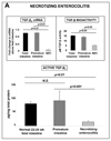
TGF-β2 expression is decreased in intestinal tissues samples resected from patients with NEC: TGF-β2 expression in tissue samples of NEC (n=8) was lower than in the mid-gestation fetal (n=6) and preterm neonatal intestine (n=5). Data show measurements by ELISA (means ± SEM). Insets show TGF-β2 mRNA expression (top left) and TGF-β bioactivity (top right). Groups were compared by Kruskall-Wallis H-test.
Fig. 6
Fig. 6
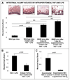
TGF-β protects mouse pups against NEC-like intestinal injury: (A) Loss of TGF-β signaling in mice worsened NEC-like intestinal injury induced by intraperitoneal administration PAF and LPS. Bar diagram (means ± SEM) shows the severity of mucosal injury on a 5-point scale in wild type mice, transgenic DNIIR mice with a partial deficiency of TGF-β signaling (3 days of zinc supplementation) and DNIIR mice with complete disruption of TGF-β signaling (7 days of zinc supplementation); n =18 mice per group. Injury scores in DNIIR mice treated with zinc for 3 or 7 days but not treated with PAF and LPS were similar to WT controls (0.24 ±0.11 and 0.26±0.20, respectively; not depicted in the bar diagram). Groups were compared by Kruskall-Wallis H-test. (B) Enteral administration of TGF-β2 prior to PAF and LPS administration in mouse pups reduced the severity of NEC-like intestinal injury; n =18 mice per group, comparison by Mann-Whitney U-test. (C) Enteral administration of TGF-β2 once a day in formula-fed mouse pups reduced the severity of intestinal injury induced by hypoxic stress; n=18 mice per group, comparison by Mann-Whitney U-test.
Fig. 7
Fig. 7
Incomplete development of macrophage tolerance to bacterial products predisposes the preterm intestine to NEC. In the mature intestine (schematic representation on the left), epithelial and stromal cell-derived TGF-β attenuates the inflammatory responses of intestinal macrophages to luminal bacteria or their products. In contrast, in the premature infant (right), the inflammatory responses of intestinal macrophages remain intact because TGF-β expression, and therefore, mucosal tolerance to bacterial products, are deficient. Bacterial products trigger an intense inflammatory reaction, causing widespread tissue damage. Enteral supplementation of recombinant TGF-β2 is a potential therapeutic strategy to prevent NEC in neonates.

Similar articles

Cited by

References

    1. Frost BL, Jilling T, Caplan MS. The importance of pro-inflammatory signaling in neonatal necrotizing enterocolitis. Semin Perinatol. 2008;32:100–106. - PMC - PubMed
    1. Hsueh W, Caplan MS, Tan X, MacKendrick W, Gonzalez-Crussi F. Necrotizing enterocolitis of the newborn: pathogenetic concepts in perspective. Pediatr Dev Pathol. 1998;1:2–16. - PMC - PubMed
    1. Bury RG, Tudehope D. Enteral antibiotics for preventing necrotizing enterocolitis in low birthweight or preterm infants. Cochrane Database Syst Rev. 2001 CD000405. - PubMed
    1. Neu J. Necrotizing enterocolitis: the search for a unifying pathogenic theory leading to prevention. Pediatr Clin North Am. 1996;43:409–432. - PMC - PubMed
    1. Smythies LE, Maheshwari A, Clements R, Eckhoff D, Novak L, Vu HL, Mosteller-Barnum LM, Sellers M, Smith PD. Mucosal IL-8 and TGF-beta recruit blood monocytes: evidence for cross-talk between the lamina propria stroma and myeloid cells. J Leukoc Biol. 2006;80:492–496. - PubMed

Publication types

LinkOut - more resources