. 2011 Jan 5;363(2):210-20.
doi: 10.1016/j.jim.2010.09.022. Epub 2010 Sep 24.

A flow cytometry-based strategy to identify and express IgM from VH1-69+ clonal peripheral B cells

Affiliations

A flow cytometry-based strategy to identify and express IgM from VH1-69+ clonal peripheral B cells

Edgar D Charles et al. J Immunol Methods. .

Abstract

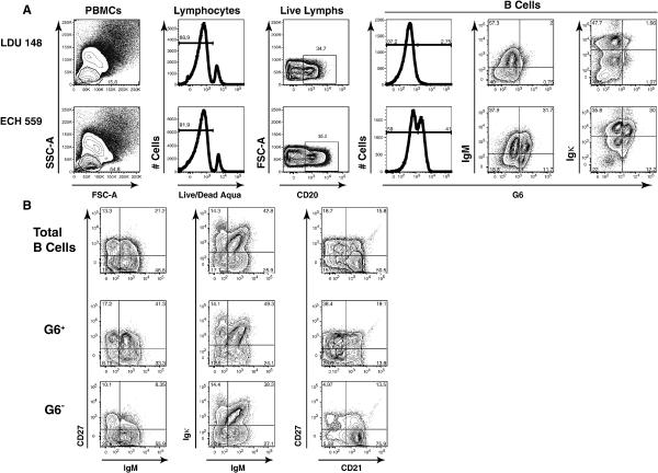
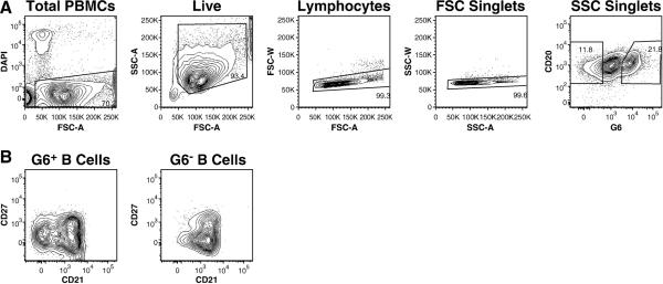
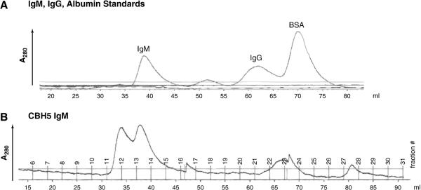
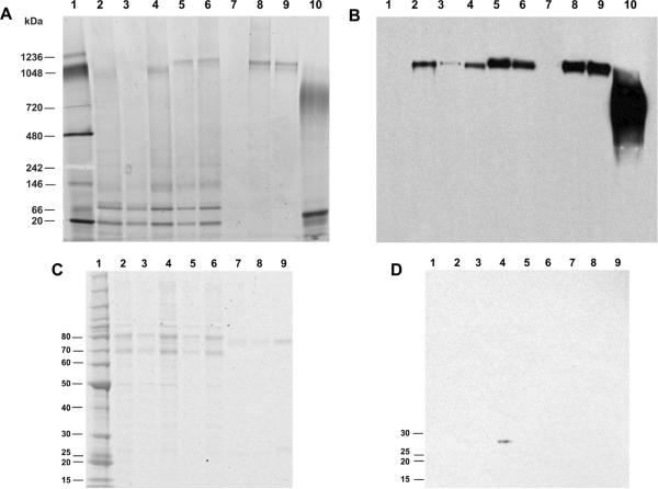
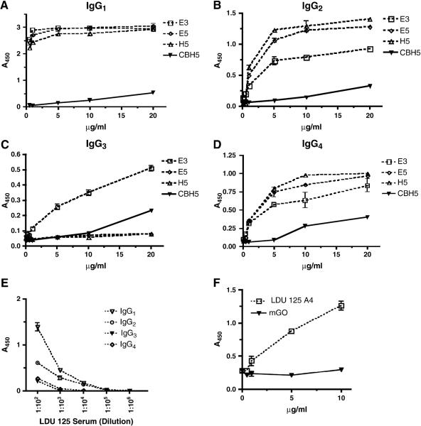
Pathologic rheumatoid factor (RF) levels are hallmarks of several human diseases. Production of monoclonal RF in vitro is essential for studies of the antigenic specificities of RF, as well as for a dissection of the mechanisms of aberrant RF+ B cell activation. We have expanded upon previous methods to develop a flow cytometry-based method to efficiently clone monoclonal antibodies (mAbs) from humans with expansions of RF-like, immunoglobulin heavy chain variable region (IgVH) 1-69 gene segment-containing B cells. The cloned variable regions are expressed as IgM and produced during culture at concentrations between 5 and 20 μg/ml. Using this system, we show that clonal Igs from patients with HCV-related mixed cryoglobulinemia, when expressed as IgM, have RF activity. We anticipate that this system will be useful for the cloning and expression of mAbs partially encoded by VH1-69 and for determination of the reactivity patterns of polyspecific, low-affinity IgMs of human pathogenic importance.


Figures

Figure 1
Figure 1. The G6 mAb identifies an expanded clonal B cell population
Flow cytometric analyses of a representative HCVMC (LDU 148) patient and a symptomatic HCV+MC+ patient with elevated serum rheumatoid factor (702 IU/ml) (ECH 559) are shown. Lymphocytes were identified by forward and side light scatter characteristics. After exclusion of dead cells, 2.8% and 41% of CD20+ B cells from LDU 148 and ECH 559, respectively, were G6+. The G6+ B cells from ECH 559 are predominantly IgM+ and Igκ+ (A). Depiction of total B cells and G6+ and G6 B cell subsets from subject ECH 559 (B). Compared to total B and G6 B cells, the G6+ B cell subset is enriched for IgM+CD27+ and IgM+κ+ B cells. The G6+ B cell subset is enriched for CD21low B cells, the majority of which are CD27+.
Figure 2
Figure 2. Sorting strategy for single-cell Ig RT-PCR
PBMCs from an HCV+MC+ patient with a monoclonal IgMκ+ B cell expansion (LDU 125) are shown. Lymphocytes were identified by forward and side light scatter characteristics. After exclusion of dead cells and doublets, G6+ CD20+ B cells were identified (A) and singly-sorted into individual wells of a 96-well microplate. CD27+/−/CD21high/low G6+ and G6 B cell subsets (B) could additionally be bulk-sorted for downstream functional analyses.
Figure 3
Figure 3. Gel filtration chromatography of expressed IgM
As sizing standards, 500 μg each of human IgMλ from a patient with multiple myeloma, human IgG1, or bovine serum albumin were separated on a HiPrep 16/60 Sephacryl S-300 HR chromatography column. A280 nm of the eluate is depicted as a function of eluate volume. The horizontal lines represent conductivity and pH (A). CBH5 IgM (500 μg) was similarly applied to the column, and 3 ml elution fractions were collected (B).
Figure 4
Figure 4. SDS-PAGE Analysis of expressed IgM
Samples run on Native PAGE 3–12% Bis-Tris gels and either stained with Sypro Ruby (A) or electroblotted to nitrocellulose and probed with HRP-conjugated anti-human IgM (B). Lane 1: NativeMark molecular weight marker; lane 2: mGO IgM transfectant supernatant (7.5 μl); lane 3: 1403 IgM transfectant (293T cells without J chain) supernatant (7.5 μl); lane 4: mGO IgM concentrated with a 100 kDa spin filter (100 ng); lane 5: 1403 IgM (produced in 293T cells without J chain) concentrated with a 100 kDa spin filter (100 ng); lane 6: 1403 IgM concentrated with a 100 kDa spin filter (100 ng); lanes 7–9: fractions 11, 12 and 13, respectively, of gel-filtered 1403 IgM concentrated with a 100 kDa spin filter (7.5 μl each); lane 10: IgM from a patient with multiple myeloma (1 μg). Reduced samples run on 4–20% Bis-Tris gels and stained with either Sypro Ruby (C) or electroblotted to nitrocellulose and probed with mouse anti-human IgJ and HRP-conjugated rabbit anti-mouse IgG (D). Lane 1: Benchmark molecular weight marker; lane 2: 773 IgM transfectant supernatant (7.5 ul); lane 3: 1403 IgM transfectant (293T cells without J chain) supernatant (7.5 ul); lane 4: mGO IgM concentrated with a 100 kDa spin column (100 ng); lane 5: 1403 IgM (produced in 293T cells without J chain) concentrated with a 100 kDa spin filter (100 ng); lane 6: 1403 IgM concentrated with a 100 kDa spin filter (100 ng); lanes 7–8: fractions 12 and 13, respectively, of gel-filtered 1403 IgM concentrated with a 100 kDa spin filter (7.5 μl each); lane 10: IgM from a patient with multiple myeloma (100 ng).
Figure 5
Figure 5. RF ELISA of mAbs
Assessment of activities of cloned IgM towards IgG isotypes. Microwells were coated with 1 μg/well monoclonal human IgG1λ, IgG2λ, IgG3λ, or IgG4λ. After blocking, serial dilutions of IgM mAbs were added. HRP-labeled goat anti-human Igμ was used as the detection Ab. E3, E5, and H5: IgM mAbs from HCV+MC+ subject LDU 125; CBH5: RF negative control, VH1-69-encoded IgM mAb (A–D). Comparison of reactivities of LDU 125 serum towards IgG isotypes (E). Recognition of cloned IgM by G6 mAb. Microwells were coated with anti-human IgM and after blocking, 0.5–10 μg/ml LDU 125 A4 IgM or mGO were added, followed by detection with G6 mAb (1:5000) and rabbit anti-mouse IgG1 (1:5000) (F).

Similar articles

Cited by

References

    1. National Center for Biotechnology Information GenBank. [Accessed October 1, 2009]. http://ncbi.nlm.nih.gov/Genbank.
    1. Bende RJ, Aarts WM, Riedl RG, de Jong D, Pals ST, van Noesel CJ. Among B cell non-Hodgkin's lymphomas, MALT lymphomas express a unique antibody repertoire with frequent rheumatoid factor reactivity. J Exp Med. 2005;201:1229–41. - PMC - PubMed
    1. Bonagura VR, Agostino N, Borretzen M, Thompson KM, Natvig JB, Morrison SL. Mapping IgG epitopes bound by rheumatoid factors from immunized controls identifies disease-specific rheumatoid factors produced by patients with rheumatoid arthritis. J Immunol. 1998;160:2496–505. - PubMed
    1. Brown CM, Plater-Zyberk C, Mageed RA, Jefferis R, Maini RN. Analysis of immunoglobulins secreted by hybridomas derived from rheumatoid synovia. Clin Exp Immunol. 1990;80:366–72. - PMC - PubMed
    1. Carbonari M, Caprini E, Tedesco T, Mazzetta F, Tocco V, Casato M, Russo G, Fiorilli M. Hepatitis C virus drives the unconstrained monoclonal expansion of VH1-69-expressing memory B cells in type II cryoglobulinemia: a model of infection-driven lymphomagenesis. J Immunol. 2005;174:6532–9. - PubMed

Publication types

MeSH terms

Associated data